 As a reminder WHY we are doing this:
What are microglia:
Microglia comprise about 10% of the cell population of the brain and form the main first immune defense of the CNS. They are phagocytic, cytotoxic, present antigens and they can promote repair after injury (Simard 2007).
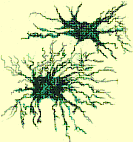
Microglia in the brain display normally a quiescent state in which phagocytosis, immune response and migration are downregulated and the microglia show a ramified form with long processes (Davalos 2005; Nimmerjahn 2005).
Microglia react to lesions by proliferation. They migrate actively towards amyloid-β plaques in 1-2 days (Meyer-Luehmann 2008) and take on an amoeboid morphology (Kreutzberg 1996; Stence 2001)
Microglia are thought to originate from a special progenitor cell during embryogenesis and that they are replenished during life by myeloid progenitors migrating from the bone marrow which invade the brain and mature to microglia (Lawson 1992; Rezaie 2002; Chan 2007;
Simard 2007). In fact it has been shown that bone marrow cells can be differentiated to microglia (Davoust 2006).
What role do microglia play in age:
The incidence of Alzheimer and autoimmune diseases in age has been hypothesized to result from non-removed debris (Streit 2006). The common hypothesis points to a disregulation of old microglia and holds that overreactive and constantly inflamed microglia might damage neuronal tissue (McGeer 1995). Newer findings suggest that microglia associated with tau pathologies are senescent instead of reactive and unable to perform function (Streit 2009). Microglia loose their ability to recognize and digest debris during age (Stolzing 2006). However studies involving complete microglia ablation for short periods did not result in increased plaque formation or neuritic dystrophy (Grathwohl 2009).
Replenishment of microglia during lifetime has been suggested to occur locally in the brain and by invasion of bone marrow progenitor cells (Streit 2006). Bone marrow transplantation has routinely resulted in bone marrow derived microglia in the brain (Rodriguez 2007) and
intravenously injected hematopoietic stem cells have migrated to the brain, differentiated into microglia and reduced infarct. Newer findings however show the necessity of irradiation to obtain sufficient numbers of invading cells (Ajami 2007; Mildner 2007). This was suspected to be due to irradiation either raising the permeability of the blood brain barrier or to provide signals strongly enhancing proliferation. This complements the view that a possible replenishment throughout age both by local proliferation and by invasion of progenitors do not suffice to prevent the slow deterioration of microglia cell population and function in age (Stolzing 2006; Streit 2006; Flanary 2007; Sierra 2007; Sawada 2008).
Prospects and pitfalls of a microglia therapy:
Both the hypothesis of disregulated, constantly inflamed microglia and the hypothesis of microglia senescence and non sufficient replenishment suggest the introduction of functional microglia as one possible solution.
The hypothesis of bystander damage by microglia (McGeer 1995) shows that the formation of massive plaques might only be a much later appearing symptom of the functional loss of the microglia population. In this view the actual Alzheimer disease starts already with the
accumulation of relatively small amounts that act chronically inflammatory and is at the time of massive plaque formation already long at work. While dysfunctional microglia can not easily be removed from the brain the introduction of functional microglia might remove the
causes for inflammation.
The hypothesis of microglia senescence and the association of dysfunctional, senescent microglia with plaques call even more readily for a cell therapy of microglia in age. In view of both hypotheses, a treatment of the symptoms, for example through down regulation of the inflammatory response, does not remove the underlying causes of aging. Even a degradation of plaques if not properly digested by dysfunctional microglia will not enhance Alzheimer condition. Therefore function of microglia has to be restored. Experiments on the influence of irradiation in bone marrow transplantation have shown that invasion of cells in absence of irradiation is low (Ajami 2007; Mildner 2007). Studies of non ablated, syngeneic conditions have however shown that massive amounts of cells are necessary to archive comparable levels of chimerism in bone marrow (Ramshaw 1995). This points to a competition for stem cells niches which is alleviated by irradiation (Colvin 2004). In age, on the other hand, the permeability of the blood brain barrier increases which has also been linked to Alzheimer (Farrall 2009; Valle 2009).
We have worked extensively on the aging and functional loss of the microglia cell population (Stolzing 2006). I optimized differentiation of microglia from bone marrow and showed - to my knowledge for the first time - the actual functional capacity of bone marrow derived microglia
(In progress of being published). Furthermore I could show that my bone marrow derived microglia invade brain slices in vitro.
Based on the hypothesis that the functional loss of microglia in age is responsible for Alzheimer we propose to use our microglia derived from bone marrow stem cells as a therapy for Alzheimer's and other age-related neurodegeneration.
We propose to introduce our differentiated microglia intravenously. Migration to the brain is tracked by sex based RTPCR. These functional microglia might then remove and digest the inflammatory debris in the brain,
therefore remove the root cause of inflammation of aged microglia. Over a longer follow up period a down regulation of inflammation and onset of neurogenesis might take place. To assess such effects numbers, density and activation states of microglia, numbers and densities of neuronal progenitor cells and number, densities and size of plaques are measured using stereology.


