Selfmade microglia
Posted by
Mondey
,
19 May 2012
·
4,205 views
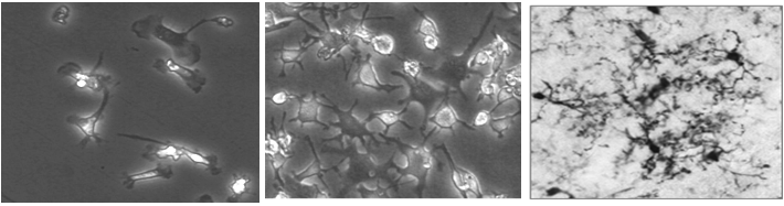 To start with the microglia replacement therapy we need lots of cells and they must resemble the original microglia. In the pictures you see microglia isolated from mice (left) or self-made microglia derived from bone marrow (in vitro differentiated).
In both cases they have basically the same morphology. In case of the self-made microglia we only have more in the dish.
You see that they look mostly ramified (spread out), however not as much as seen in brain tissue (in vivo). You can read all the details in our publications (Hinze and Stolzing, 2011; Hinze and Stolzing, 2012).