.
F U L L T I T L E : Swimming exercise stimulates IGF1/ PI3K/Akt and AMPK/SIRT1/PGC1α survival signaling to suppress apoptosis and inflammation in aging hippocampus.
O P E N A C C E S S S O U R C E : AgING
Abstract
Hippocampus is one of the most vulnerable brain regions in terms of age-related pathological change. Exercise is presumed to delay the aging process and promote health because it seems to improve the function of most of the aging mechanisms. The purpose of this study is to evaluate the effects of swimming exercise training on brain inflammation, apoptotic and survival pathways in the hippocampus of D-galactose-induced aging in SD rats. The rats were allocated to the following groups: (1) control; (2) swimming exercise; (3) induced-aging by injecting D-galactose; (4) induced-aging rats with swimming exercise. The longevity-related AMPK/SIRT1/PGC-1α signaling pathway and brain IGF1/PI3K/Akt survival pathway were significantly reduced in D-galactose-induced aging group compared to non-aging control group and increased after exercise training. The inflammation pathway markers were over-expressed in induced-aging hippocampus, exercise significantly inhibited the inflammatory signaling activity. Fas-dependent and mitochondrial-dependent apoptotic pathways were significantly increased in the induced-aging group relative to the control group whereas they were decreased in the aging-exercise group. This study demonstrated that swimming exercise not only reduced aging-induced brain apoptosis and inflammatory signaling activity, but also enhanced the survival pathways in the hippocampus, which provides one of the new beneficial effects for exercise training in aging brain.
Introduction
Aging is a gradual process, the accumulation of molecular and cellular damage throughout life leads to a wide range of age-related disorders such as cardiovascular dysfunction and neurodegenerative diseases. Studies have demonstrated that increased levels of reactive oxygen species (ROS) are involved in the detrimental effect of aging-induced cell damage [1]. Natural aging was experimentally modeled by chronic administration of D-galactose in rodents, as it reacts with amino acids in proteins resulting in formation of advanced glycation end-products(AGEs) binding of AGEs to their receptors causes ROS production and AGEs, which are elevated in aging, neurodegeneration and inflammation [2]. Numerous studies indicated that physical exercise exerts neuroprotective effects and increases cognitive activity [3], ameliorates neurological impairments in several neurodegenerative diseases [4] and reduces age-related neuronal loss [5]. Physical exercise is known to have positive effects on the central nervous system via a number of mechanisms, including increases neurotrophic factors production such as VEGF, IGF1, NGF and BDNF that lead to improved efficiency of the vascular system and trigger neuroplastic mechanism in the brain [6–9].
SIRT1, a type III histone/protein deacetylase, is considered a novel anti-aging protein involved in the regulation of variety cell signaling pathways, including apoptosis, cellular senescence, proliferation, oxidative stress, energy metabolism, inflammation and DNA damage response [10]. SIRT1 is abundantly expressed in several regions of the brain, including the hypothalamus, cortex, striatum and hippocampus [11]. A neuroprotective effect of SIRT1 has been reported for both acute and chronic neurological diseases [12]. SIRT1 regulates p53, NF-κB, FOXO family, PGC-1α, and other transcription factors via deacetylation [12]. Caloric restriction and resveratrol intake slow the aging process and promote successful brain aging, with SIRT1 being a key regulator [12]. The AMP-activated protein kinase (AMPK) and SIRT1 are evolutionary conserved partners which have similar functions in metabolism, energy balance and cellular survival [13]. Recently, the effect of exercise on SIRT1 expression and activity has started to be investigated. In aged rats, SIRT1 activity has been reported to increase after a 6 weeks treadmill training program in heart and adipose tissue [14]. In contrast, in rat cerebellum SIRT1 activity was decreased with aging and it was not increased by mild exercise [15]. In humans, three days of cycling caused increases in mRNA of SIRT1 and PGC-1α in skeletal muscle [16] and marathon running increased SIRT1 and other anti-apoptotic gene expression in peripheral blood mononuclear cells [17]. While most of the effects of exercise on SIRT1-dependent pathways have been described in peripheral tissues, these observations allow hypothesizing that some of the beneficial neurophysiological effects of exercise could be mediated by the modulation of SIRT1 activity in the brain.
Several evidences have indicated that IGF1 plays a crucial role in protection of neurons and low IGF1 levels are associated with brain aging [18]. Activation of IGF1 is beneficial to improve neuronal survival during brain aging [19]. These actions are mediated by IGF1 receptors widely distributed in the brain [20]. The cell survival property of IGF1R signaling is mediated by the activation of the phosphatidylinositol 3-kinase (PI3K) and protein kinase B (Akt) [21, 22]. Phosphorylated Akt serves as a pro-survival signaling pathway by affecting the Bcl-2 family [23]. Bcl-2 family can be divided into pro-apoptotic and pro-survival subgroups. Bcl-2 and Bcl-xL are pro-survival proteins that prevent the activation of downstream apoptotic signaling. However, IGF1R and Bcl-2 family associated pro-survival pathway in aging brain is still not completely understood.
Implicated inflammation was found in aging process and neurodegenerative diseases [24]. It was suggested that alterations in pro-inflammatory cytokines levels, such as tumor-necrosis factor alpha (TNFα), which can potently be induced following brain injury and promote neuroinflammation and neurodegeneration [25]. TNF-α pathway is mediated through two distinct cell surface receptors: TNFR1 and TNFR2. Only TNFR1 contains a cytoplasmic death domain and may directly induce apoptosis [26]. NF-κB also regulates expression of various neurotrophic proteins implicated in neuronal development, function and survival factors [27]. Genes known to be regulated by NF-κB include cyclooxygenase-2 (COX-2) and inducible nitric oxide synthase (iNOS) [27]. Excessive NO generation by iNOS may be harmful to the brain.
Many studies have demonstrated that apoptosis may contribute to the loss of neuronal cells in neuropathy and is regarded as a predictor of adverse outcomes in subjects with age-related neurodegenerative diseases [28]. Apoptosis has two main pathways, extrinsic Fas-dependent and intrinsic mitochondrial-dependent apoptotic pathways. The extrinsic Fas-dependent pathway is initiated by binding the Fas ligand to the Fas receptor. After ligand binding, Fas-receptor oligomerization results in the recruitment of the Fas associated death domain (FADD) adaptor protein and the activation of caspase 8, which is upstream of caspase 3 that is responsible for DNA-cleavage action [28]. Mitochondrial-dependent apoptotic pathway is mediated by internal factors, such as free radicals. In this pathway, mitochondria play an important role in apoptosis by releasing cytochrome c and activating caspase 9, which activates caspase 3. Our recent study indicated that diabetes and exposure of side stream smoke cause Fas receptor-dependent and mitochondrial-dependent apoptosis in animal brains [29, 30].
In this study, we will determine the expression patterns of SIRT1 and related longevity genes in control and induced aging brain of rats, and investigated whether swimming exercise affected their expression. Furthermore, we want to understand whether the IGF1R signaling and pro-survival Bcl-2 family associated pathways in induced- aging brain are worse than control rats and whether these components can be improved by swimming exercise program. We hypothesized that aging may predispose to more impaired neuronal IGF1R/Akt and pro-survival Bcl-2 family pathways, as well as swimming exercise may enhance neuronal IGF1R/Akt survival and AMPK/SIRT1 pathways and prevent apoptotic activity in aging hippocampus.
Results
Pathohistological changes of hippocampal neurons
To investigate changes of neuronal architecture in D-galactose-induced ageing after swimming exercise, we performed a histopathological analysis of hippocampal tissue stained with hematoxylin and eosin (HE). Figure 1 shows representative micrographs of HE staining in the cornu ammonis 1 (CA1), cornu ammonis 3 (CA3) and dentate gyrus (DG) regions of hippocampus for each group. D-galactose-induced aging showed the tendency to decrease the neuronal cell density, which was increased by swimming exercise treatment.
Figure 1. Representative micrographs of H&E staining in the CA1, CA3 and dentate gyrus (DG) regions of hippocampus for each group. The images of hippocampus architecture were magnified 400 times.
AMPK/SIRT1 longevity-related signaling pathway
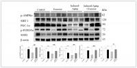
To identify the effects of D-galactose-induced aging and swimming exercise on the p-AMPK/SIRT1 longevity-related signaling pathway in rat hippocampus, western blotting was conducted. The protein levels of p-AMPK, SIRT1, PGC-1α, p-FOXO3a and SOD2 were significantly decreased in induced-aging group compared with those in the control group. However, these proteins levels in the induced-aging+exercise group were significantly increased compared with the induced-aging group (p<0.05, Figure 2). In the control group, swimming exercise mildly enhanced the expression of p-AMPK and SOD2. And swimming exercise mildly suppressed the expression of SIRT1 and PGC-1α (Figure 2).
Figure 2. Exercise training enhanced AMPK/SIRT1 anti-aging pathway in D-galactose-induced aging rat hippocampus. The representative protein levels of p-AMPKα, SIRT1, PGC-1α, p-FOXO3a, SOD2 prepared from hippocampi in the control, exercise, aging and aging with exercise rats were measured by Western blotting analysis (n=3). The protein expression folds were normalized with β-actin. * P <0.05, **P<0.01, ***P<0.001, significant differences from the control group. ##P <0.05, ###P <0.01, significant differences from the D-galactose-induced aging group.
IGFI-R/PI3K/Akt survival pathway
To investigate the correlation between longevity-related signaling molecules and the IGF1R/Akt survival pathway, we examined the protein levels of p-IGFIR, p-PI3K, p-Akt and Bcl-xL. The protein levels of p-PI3K, p-Akt and Bcl-xL in the induced-aging group were significantly lower than those in the control group, but p-IGF1-R expression was not altered. However, swimming exercise effectively enhanced the expression of those survival proteins in induced-aging+exercise group compared to induced-aging group (Figure 3). Conversely, swimming exercise alone exerted no significant effect on the expression of these survival proteins compared to control group (Figure 3).
Figure 3. Exercise training enhanced IGF1/Akt survival pathway in D-galactose-induced aging rat hippocampus. The representative protein levels of survival proteins p-IGF1 receptor (p-IGF1R), p-PI3K, p-Akt and pro-survival Bcl family of Bcl-xL prepared from hippocampi in the control, exercise, aging and aging with exercise rats were measured by Western blotting analysis (n=3). The protein expression folds were normalized with β-actin. ***P<0.001, significant differences from the control group. ##P <0.05, ###P <0.01, significant differences from the D-galactose-induced aging group.
Stress-related inflammatory proteins
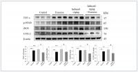
To verify the effects of swimming exercise and aging on inflammatory proteins, we examined the protein levels of TNFα, p-NFκB, COX-2 and iNOS for each group. It was observed a marked increase on hippocampal TNFα, p-NFκB, COX-2 and iNOS levels in the induced-aging group when compared to the control group. Swimming exercise attenuated the aging induced increase in the expression of these proteins (Figure 4). Conversely, swimming exercise significantly increased the expression of those inflammatory proteins compared to control group (Figure 4).
Figure 4. Effects of exercise training on inflammatory proteins. The representative protein levels of TNFα, p-NFκB, iNOS and COX2 prepared from hippocampi in the control, exercise, aging and aging with exercise rats were measured by Western blotting analysis (n=3). The protein expression folds were normalized with β-actin. **P<0.01, ***P<0.001, significant differences from the control group. ##P <0.05, significant differences from the D-galactose-induced aging group.
.../...
Discussion
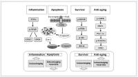
Our main findings can be summarized as follows (Figure 7): (1) The Fas dependent, mitochondrial-dependent apoptotic pathways and inflammatory proteins exhibited increased levels with a reduced IGFI-R/PI3K/Akt and AMPK/SIRT1 survival pathways in the hippocampus of D-galactose induced aging rats. (2) Swimming exercise not only enhanced IGFI-R/PI3K/Akt and AMPK/SIRT1 survival pathways but also reduced the apoptotic and inflammatory pathways in hippocampus from D-galactose induced aging rats. (3) Swimming exercise modulated the apoptotic and inflammatory pathways in an induced aging-dependent manner, swimming exercise was able to reverse the increases of the apoptotic and inflammatory pathways activity induced by aging process, while increasing the levels of apoptotic and inflammatory protein in control hippocampus.
Figure 7. Our hypothesis that exercise training suppressed inflammatory and apoptotic pathways and enhanced IGF1R/PI3K/Akt survival and anti-aging AMPK/SIRT1/PGC-1α associated pathways in the hippocampus of D-galactose induced aging rats. These aging attenuated IGF1R related PI3K/Akt survival pathway and AMPK/SIRT1/PGC-1α pathway were improved after exercise training. Activated inflammatory and apoptotic activity were prevented after exercise training.
.../...
.


