.
O P E N A C C E S S S O U R C E : Mechanisms of ageing and Development
Highlights
• Mild and repeated doses of UVB induce senescence of human epidermal melanocytes.
• UVB-irradiated melanocytes show impaired proteasome activity and increased autophagy.
• UVB irradiation induces melanogenesis in melanocytes.
• Photoaged fibroblasts secrete factors that boost melanogenesis of melanocytes.
Abstract
Ultraviolet (UV) light is known to potentially damage human skin and accelerate the skin aging process. Upon UVB exposure, melanocytes execute skin protection by increasing melanin production. Senescent cells, including senescent melanocytes, are known to accumulate in aged skin and contribute to the age-associated decline of tissue function. However, melanocyte senescence is still insufficiently explored. Here we describe a new model to investigate mechanisms of UVB-induced senescence in melanocytes and its role in photoaging. Exposure to mild and repeated doses of UVB directly influenced melanocyte proliferation, morphology and ploidy. We confirmed UVB-induced senescence with increased senescence-associated β-galactosidase positivity and changed expression of several senescence markers, including p21, p53 and Lamin B1. UVB irradiation impaired proteasome and increased autophagic activity in melanocytes, while expanding intracellular melanin content. In addition, using a co-culture system, we could confirm that senescence-associated secretory phenotype components secreted by senescent fibroblasts modulated melanogenesis. In conclusion, our new model serves as an important tool to explore UVB-induced melanocyte senescence and its involvement in photoaging and skin pigmentation.
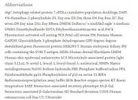
Abbreviations

1. Introduction
The skin is the largest organ of the human body and performs several functions, including protection against damage caused by environmental factors (Prieux et al., 2020). Skin aging is caused by two distinct, but overlapping, mechanisms – intrinsic and extrinsic aging. Intrinsic aging affects not only the skin, but all tissues and organs of the body and is understood as time-depending and influenced by genetic background (Abd El-Aal et al., 2012; Cavinato et al., 2017). In addition, skin is continuously exposed to environmental factors, which contribute to the process of extrinsic skin aging. Among the factors that cause damage to the skin, UV radiation is considered the most harmful (Cavinato, 2020; Krutmann et al., 2017). UVB rays are known to directly damage DNA, to promote the accumulation of reactive oxygen species (ROS) and subsequent oxidation of proteins and lipids (Greussing et al., 2013).
Cellular senescence is a state of proliferative arrest characterized by significant changes in the morphology and functionality of cellular components (López-Otín et al., 2013). Accumulation of senescent cells in different tissues contributes to the decline of functions characteristic of the aging process (Baker et al., 2016; Tominaga, 2015). Specifically in the skin, senescent fibroblasts contribute to aging by secretion of extracellular matrix degrading matrix metalloproteases (MMPs) and other senescence-associated secretory phenotype (SASP) components (Toutfaire et al., 2017; Waldera Lupa et al., 2015; Wang and Dreesen, 2018). Senescence of melanocytes has been insufficiently explored but recent studies have reported that senescent melanocytes accumulate in human skin where they contribute to the aging process by impairing proliferation of the neighboring keratinocytes (Victorelli et al., 2019; Waaijer et al., 2016).
A decisive characteristic of skin aging is the accumulation of oxidized and damaged proteins, which leads to an impaired cellular protein homeostasis (Cavinato and Jansen-Dürr, 2017). In a previous publication, we have demonstrated that damaged proteasome activity is compensated by increased autophagy in a model of UVB-induced senescence of fibroblasts and that these events are essential for the establishment of the senescence phenotype (Cavinato et al., 2016). Skin aging-associated proteostatic changes also occur in keratinocytes (Eckhart et al., 2019). Autophagy is closely connected to the regulation of synthesis and removal of melanosomes and defects in either one of these mechanisms potentially causes pigmentary disorders (Ho and Ganesan, 2011; Wang et al., 2019b). The exact role of proteasome and autophagic activity in melanocytes - especially after UV-exposure of the cells - is not yet understood.
Age spots typically occur in chronically sun-exposed skin and the appearance of this aberrant pigmentation has been associated with irregularities in melanocyte distribution, enhanced melanogenic signalling and decreased melanosome removal (Barysch et al., 2019; Choi et al., 2017; Haddad et al., 1998). Recently, it has been demonstrated that the occurrence of age spots is not solely influenced by changes in melanocytes, but by the deregulated secretion of molecules produced by photoaged keratinocytes and fibroblasts emphasizing the complexity of underlying factors driving photoaging and the development of senile lentigines (Bellei and Picardo, 2020). However, there is still a big gap in our understanding of the underlying mechanisms by which senescent cells contribute to the photoaging of the skin. Thus, the establishment of models that reflect these processes is increasingly necessary.
Here, we introduce a new model to study melanocyte senescence during the process of photoaging. Using this model, we investigated the direct influence of UVB irradiation on melanocyte proteasome and autophagic activity in cell monolayers. In addition, we examined the potential of a previously established UVB-induced senescence model of dermal fibroblasts (Cavinato et al., 2016; Greussing et al., 2013) to influence aspects of melanocyte's biology in a co-culture system.
2. Material and methods
.../...
3. Results
3.1. UVB induces changes in proliferation, morphology and ploidy of human epidermal melanocytes
Previous work of our group has demonstrated that mild and repeated doses of UVB lead to stress-induced premature senescence of human dermal fibroblasts (Cavinato et al., 2016; Greussing et al., 2013). Using the same experimental approach, in which cells are subjected to UVB-irradiation twice a day for a period of four consecutive days, we investigated the direct effect of UVB on melanocytes (Fig. 1 A). Initially, to ensure that the melanocyte cultures would not be contaminated by fibroblasts, characterization of melanocytes derived from newborn foreskin was performed by immunofluorescence to detect the presence of the melanocyte marker Melan-A (Sup. Fig. 1). Then, to establish the proper conditions through which UVB induces senescence, melanocytes were submitted to different dosages of UVB and monitored for cell proliferation (Sup. Fig. 2). Control non-irradiated cells performed around 7 population doublings (cPDLs) during the 15 days of experiment. Melanocytes irradiated with 0.075 and 0.1 J/cm2 UVB displayed slight growth arrest. Treatment of cells with 0.125 J/cm2 UVB induced stronger growth arrest, while doses equal to or greater than 0.150 J/cm2 UVB led to negative cPDL numbers, indicating cell death (Fig. 1 B, S2). Based on these results, the dosage of 0.125 J/cm2 was defined as standard condition, which was used to perform all the other UVB irradiations of melanocytes described in this work.

Fig. 1. UVB affects proliferation, morphology and ploidy of human epidermal melanocytes. (A) Graphic representation of the experimental model in which cells are subjected to UVB-irradiation twice a day for a period of four consecutive days and maintained in culture for further 11 days. (B-E) Melanocytes were submitted to 0.125 J/cm2 UVB as described and monitored for cell growth (B) cell morphology (C-D) and percentage of multinucleated cells (E). (B) Growth curve of UVB-irradiated and control melanocytes. © Representative pictures of UVB-irradiated melanocytes and the respective controls obtained on day 4, 9 and 15 of the experiment. Black arrows indicate the accumulation of melanin in UVB treated melanocytes. Scale bar 50 μm. (D) Analysis of cell surface area. (E) Percentage of multinucleated cells on D4, D9 and D15. Results are presented as mean values ± SD of at least three independent experiments. *<0.05; **<0.01; ***<0.001 and n.s. no statistical significance.
Cellular senescence is characterized by cell-cycle arrest and alterations in cell shape and metabolism (Greussing et al., 2013). In this way, cell morphology and number of nuclei per cell of UVB-irradiated and control melanocytes were monitored in order to further characterize the changes caused by UVB (Fig. 1). Analysis of cell surface area revealed that irradiated cells showed a significant increase in size compared to non-irradiated cells (Fig. 1C–D). The observed increase in size coincided with the growth arrest caused by the irradiation (Fig. 1 B). In addition, accumulation of cytoplasmic brown granules, indicative of increased melanin synthesis, was observed in some UVB-irradiated melanocytes (Fig. 1 B, arrows).
In certain cell types, senescence is associated with polyploidy and multinucleation due to cell cycle arrest (Akakura et al., 2010). With regard to melanocytes specifically, the appearance of multinucleated cells is related to both senescence and nevi formation (Leikam et al., 2015). By counting the number of multinucleated cells in UVB and control melanocytes we found that UVB treatment increased percentage of cells with two or more nuclei already on D4 (Fig. 1E). On D9, more than 30 % of UVB irradiated melanocytes presented more than one nucleus and on D15 this percentage was slightly decreased but still around five times higher than observed in control cells (Fig. 1E).
Cytochemistry for the activity of SA-β-Gal and analysis of the regulation of senescence-associated proteins were used to confirm senescence induction by UVB irradiation of melanocytes (Fig. 2). UVB induced the accumulation of SA-β-Gal positive cells (Fig. 2A–B) and a strong but transient phosphorylation of p53 on serine 15 (pp53), most notable at D4, i.e. after application of the last stress, indicative of p53 activation (Fig. 2C–E). In addition, the overall levels of p53 protein were also increased and remained higher than the ones from control cells throughout the experiment. Activation of p53 also resulted in the upregulation of its downstream effector p21waf1 (Fig. 2C–E). Expression of the nuclear lamina protein Lamin B1 was initially slightly increased in UVB-irradiated cells. However, on D9, coinciding with the increased activity of SA-β-Gal and expression of other senescence markers, levels of Lamin B1 were significantly lower in irradiated melanocytes than in control cells (Fig. 2C–E). Altogether these results demonstrate that mild and repeated doses of UVB induce senescence of human melanocytes.

Fig. 2. UVB induces senescence in human epidermal melanocytes. (A) Representative pictures of control and UVB-irradiated melanocytes stained for SA-β-Gal on D9 of the experiment. Scale bar 50 μm. (B) Percentage of SA-β-Gal positive melanocytes. For each group at least 300 cells were analyzed. © p21, pp53 (serin 15), p53 and Lamin B1 protein expression of UVB-irradiated and the respective control cells were analyzed by western blot on days 4 and 9 of the experiment. Representative pictures are shown. Lysates from HDFs passage 35 and HDFs treated with 33 μM Cisplatin were used as positive controls. GAPDH was used as a protein loading control. (D-E)single bond Analysis of relative intensity of the Western blot on days 4 (D) and 9 (E) was performed in ImageJ software. Results are presented as mean values ± SD of at least 3 independent experiments. *<0.05; **<0.01; ***<0.001 and n.s. no statistical significance.
3.2. UVB irradiation impairs proteasome activity and increases autophagy in melanocytes
Next, we wanted to investigate if mechanisms of protein quality control would be affected during the process of UVB-induced senescence of melanocytes. To monitor proteasome activity, we generated a reporter cell line consisting of melanocytes expressing a GFP-degron protein. Under physiological conditions, GFP-degron expressing cells show low GFP fluorescence because the destabilized protein is degraded by proteasomes. In contrast, when proteasome activity is blocked, GFP-degron protein cannot be efficiently degraded and accumulates in the cytoplasm leading to increased green fluorescence (Greussing et al., 2012) (Fig. 3 A). GFP-degron expressing melanocytes were submitted to UVB as described, and proteasome activity was monitored by FACS analysis. Non-transfected cells were used as negative control. As positive controls GFP-degron melanocytes were treated with the proteasome inhibitor N-acetyl-L-leucyl-L-leucyl-leucyl-L-norleucinal (LLnL). UVB treatment led to increased cytoplasmic green fluorescence in comparison to control nonirradiated cells throughout the whole experimental procedure, suggesting significantly impaired proteasome activity (Fig. 3 B).

Fig. 3. UVB impairs proteasome activity and induces autophagy in melanocytes. (A) Schematic representation of GFP-degron system to monitor proteasome activity in living cells. (B) Melanocytes were irradiated with UVB and green fluorescence resulting from proteasome inactivation was analyzed by flow cytometry. © UVB-irradiated and control melanocytes processed for immunofluorescence to label Atg7. Representative pictures are shown. Scale bar 50 μm. (D) Number of Atg7 puncta per cell was counted in ImageJ software. Results are presented as mean values ± SD of at least three independent experiments. *<0.05; **<0.01; ***<0.001 and n.s. no statistical significance (For interpretation of the references to colour in this figure legend, the reader is referred to the web version of this article).
To address a potential role of autophagy in UVB-induced senescence of melanocytes, cells were further processed for immunofluorescence to label Atg7. The Atg7 protein is involved in the conversion of the microtubule-associated protein light chain 3 (LC3-I) into its lipidated form, LC3-II and is, therefore, essential for the assembly of the autophagosomes (Zhang et al., 2015). UVB-irradiated melanocytes presented more Atg7 positive dots in comparison to control non-irradiated cells throughout the whole experiment (Fig. 3C–D). The peak of autophagy was observed on D9 when the mean number of Atg7 positive punctae per cell was four times higher in irradiated melanocytes in comparison with control cells. Taken together these results suggest that the process of UVB-induced senescence of melanocytes involves impairment of proteasome activity and exacerbated autophagic activity.
.../...
.
Edited by Engadin, 29 September 2020 - 06:45 PM.


