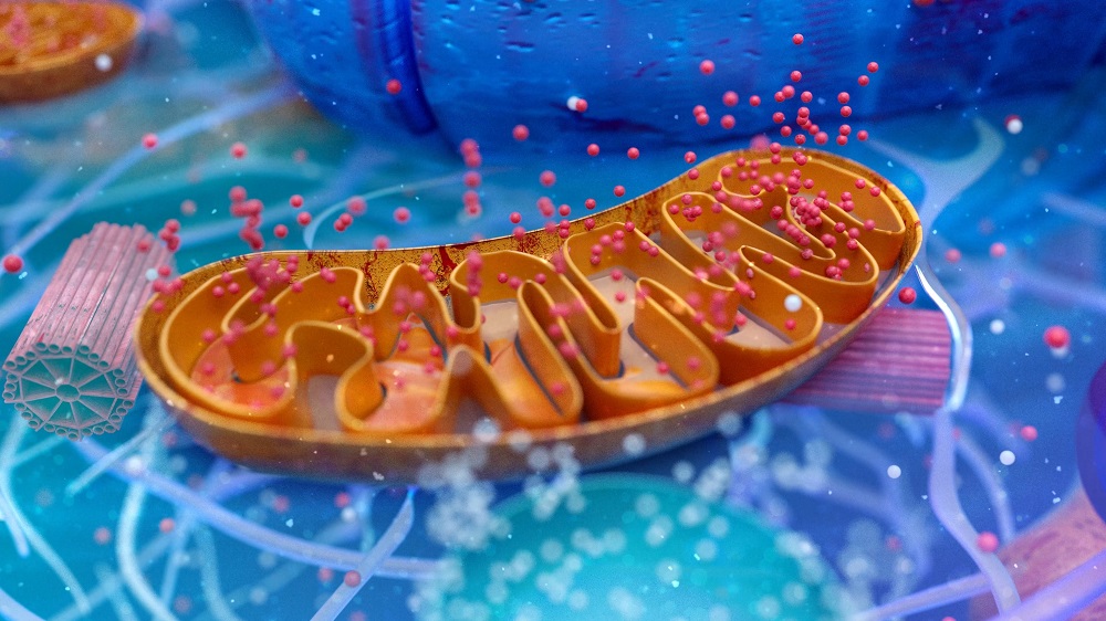
Creating induced pluripotent stem cells (iPSCs) causes mutant mitochondrial populations to change, and researchers have investigated this phenomenon more thoroughly.

How Cellular Reprogramming Affects Mitochondrial Dysfunction
#1
Posted 19 December 2024 - 05:33 PM
Being outside of the protection of the nucleus, mitochondria DNA (mtDNA) mutates at anywhere from 10 to 100 times the rate of nuclear DNA [1]. At only 16,569 base pairs, mtDNA is very compact and does not contain the extra, non-coding introns that nuclear DNA has; therefore, any mutation may have a signficant effect. If a mutation occurs to all the mitochondria in a cell, it is homoplasmy, but if it only occurs to some of them, it is heteroplasmy, and its effects vary between cell types [2]. Heteroplasmy can have severe downstream consequences, including heart problems [3].
It is well-known that within certain cell types, mitochondria with harmful mtDNA deletions outcompete mitochondria that are more beneficial for the cell [4]. However, it remains unknown precisely why this is the case. Furthermore, while mitochondrial dysfunction has plenty of age-related consequences, it has not been proven if this DNA deletion is behind some or all of them. There are also few effective ways to directly alter mitochondria in living organisms.
However, previous work has found that cellular reprogramming can change the heteroplasmy of mitochondria. Interestingly, it can go one of two ways: either the heteroplasmy becomes dominant or becomes erased entirely [5]. This has a substantial effect on stem cells, which are greatly influenced by the quality of their mitochondria [6].
All or nothing
To start their experiment, the researchers used OSKM to reprogram three distinct cell lines, one with a well-known point mutation called A3243G and the others with a frequently discussed deletion called Δ4977, although the A3243G cells had 89% of their mitochondria affected while both of the Δ4977 lines had very low percentages of this deletion. As expected, the A3243G cells had substantial problems with respiration, while the Δ4977 cells were not significantly affected.
In all cases, the cells quickly became very strongly biased for or against these mitochondrial alterations. Some of the iPSCs generated from A3243G had similar percentages to that of the original cells, while others had none whatsoever. Similarly, while the stem cell generation process initially increased the amount of Δ4977 deletions, this amount dropped very quickly in nearly all of the generated cells after only four divisions; only cells with extremely high amounts of Δ4977 retained this mutation. Cells with approximately 50% of mitochondria containing the mutation either quickly lost it or gained more of it after four divisions.
While the stem cells with large percentages of mutations did not change how they differentiated into somatic cells, there were significant effects later on. The cells with large amounts of Δ4977 grew larger but divided less than the cells without it. While these mitochondrial mutants were able to slowly differentiate into fat cells and bone cells without any visible problems, they failed to beat properly when they were differentiated into heart tissue, and critical compounds necessary for cardiac function were not found in these cells.
As expected, these Δ4977 mutants also had significant alterations in nuclear gene expression as well: genes related to fundamental metabolism, oxidative stress, small molecule transport, and the management of cholesterol were all affected. There were also trends involving such aspects of metabolism as ADP to ATP translation and the usage of NAD+.
While A3243G mutants did not have a significant difference in epigenetic age, Δ4977 mutants did. These are freshly reprogrammed cells, which should have a minimum of difference, but this difference was greater than that of iPSCs taken from 20-year-olds and iPSCs taken from 100-year-olds, according to the Horvath epigenetic clock.
These findings are of interest to some researchers looking for a supply of mitochondrially mutated cells for use in the study of mitochondrial diseases, but others may simply feel relieved. While it is not clear if these results hold true for every mitochondrial mutation, the simple process of iPSC creation appears to create either significantly affected cells that can be discarded at the clinical level or cells whose gradually accruing mitochondrial mutations have been removed with no additional intervention required. This bodes well for iPSC-based therapies, particularly those that create cardiac and similar functional tissues.
Literature
[1] Allio, R., Donega, S., Galtier, N., & Nabholz, B. (2017). Large variation in the ratio of mitochondrial to nuclear mutation rate across animals: implications for genetic diversity and the use of mitochondrial DNA as a molecular marker. Molecular biology and evolution, 34(11), 2762-2772.
[2] Picard, M., Zhang, J., Hancock, S., Derbeneva, O., Golhar, R., Golik, P., … & Wallace, D. C. (2014). Progressive increase in mtDNA 3243A> G heteroplasmy causes abrupt transcriptional reprogramming. Proceedings of the National Academy of Sciences, 111(38), E4033-E4042.
[3] Baris, O. R., Ederer, S., Neuhaus, J. F., von Kleist-Retzow, J. C., Wunderlich, C. M., Pal, M., … & Wiesner, R. J. (2015). Mosaic deficiency in mitochondrial oxidative metabolism promotes cardiac arrhythmia during aging. Cell metabolism, 21(5), 667-677.
[4] Khrapko, K., Bodyak, N., Thilly, W. G., Van Orsouw, N. J., Zhang, X., Coller, H. A., … & Wei, J. Y. (1999). Cell-by-cell scanning of whole mitochondrial genomes in aged human heart reveals a significant fraction of myocytes with clonally expanded deletions. Nucleic acids research, 27(11), 2434-2441.
[5] Wei, W., Gaffney, D. J., & Chinnery, P. F. (2021). Cell reprogramming shapes the mitochondrial DNA landscape. Nature Communications, 12(1), 5241.
[6] Chakrabarty, R. P., & Chandel, N. S. (2021). Mitochondria as signaling organelles control mammalian stem cell fate. Cell stem cell, 28(3), 394-408.
View the article at lifespan.io
#2
Posted 20 December 2024 - 02:45 PM
SOX2 encourages mitophagy. That helps.
The issue, however, is inefficient mitochondria from duff mtDNA. Likely that is heteroplasmy, but in theory it could still have the same rubbishy DNA changes to multiple copies of the mtDNA in any one mitochondrion.
Edited by johnhemming, 20 December 2024 - 02:47 PM.


