• Log in with Facebook Log in with Twitter Log In with Google      Sign In    
  • Create Account
  LongeCity
              Advocacy & Research for Unlimited Lifespans

Photo
- - - - -

Oxidative stress/glutathinone deficiency theory of alzheimer's

alzheimers glutathione tau amaloid

  • Please log in to reply
7 replies to this topic

#1 aza

  • Guest
  • 136 posts
  • 29
  • Location:aus
  • NO

Posted 04 January 2025 - 04:27 AM


Hey everyone, Im making this post because think that the oxidative stress hypothesis of alzheimer's is frequently overlooked in favor of  amyloid and tau.

I primarily looked into this to improve my nootropic stack. I'm not an expert, doctor or your mother and I'm certainly not the first to come up up with this, feedback is welcome.


I'm going to start off with the TLDR and some things i found promising: I believe that Alzheimer's is primarily the result of sirt6/glutathione deficiency which results the brain using tau to control double strand breaks without having enough resources (perhaps soluble ab42, more on that later) to effectively remove it. 


So nac + glycine (glutathione precursors, better then serine imo) + carnitine + niacinamide riboside similar to the study below would likely be helpful


https://www.ncbi.nlm.nih.gov/pmc/articles/PMC9879258/

Nac with cofactors serine, niacinamide and l-carnitine improve ADAS-Cog scores by 29% (although i think the doses are a bit much)


In addition to these i think that ergothionine would further help to normalize this via the nrf2/ARE pathway.


vitamin b12 + omega 3 are possibly synergistic in preventing brain atrophy

https://www.scienced...002916523277655


The addition of choline (or citicoline for uridine conversion) may improve synaptogenesis. In essence this part is basically the mrhappy stack.

https://pmc.ncbi.nlm...les/PMC4011061/


A valter longo style 3-5 day fasting mimicking diet is also likely to have a positive effect. It would clear out more trash while helping with neural stem cells.

https://www.ncbi.nlm...les/PMC9648488/


I also believe that vitamin k2 would help, for more then a few reasons.


Now onto the meat and potatoes, sorry in advance for the terrible formatting.


Glutathione and metal disruption
1.    Glutathione is required for proper copper metabolism and a deficiency may cause a copper deficiency in the brain as well as a build of iron due to a lack of functional ceruloplasmin. You would expect to see a intracellular copper deficiency, a buildup of extracellular copper and dysfunction iron deposition due to a lack of ceruloplasmin,
https://pubmed.ncbi....h.gov/23426973/
“depleting cellular GSH using L-buthionine-sulfoximine (BSO) caused a 50% decrease in the initial rate of (64)Cu entry in HEK293 cells and other cell types.”

https://pubmed.ncbi....h.gov/28654115/
“copper levels were substantively decreased in all AD-brain regions, to 52.8-70.2% of corresponding control values, consistent with pan-cerebral copper deficiency”

2.    Ferroptosis and iron deposition in the brain tracks strongly with Alzheimer’s and is activated via glutathione deficiency
https://www.ncbi.nlm...les/PMC9445173/
“Glutathione was found to be significantly depleted in mild cognitive impaired (P < 0.05) and Alzheimer’s disease patients (P < 0.001) as compared with healthy old participants. A significant higher

level of iron was observed in left hippocampus region for Alzheimer’s disease patients as compared with healthy old (P < 0.05) and mild cognitive impairment (P < 0.05).”


https://www.nature.c...598-022-22761-5

“The data clearly show that aggregated Aβ1-42 alone is significantly less toxic to hippocampal cells. Aggregated Aβ damages neurons, and glial cells proliferate to remove Aβ from the hippocampus. External prooxidant agents (Fe2+) or inhibition of internal antioxidant defence by BSO has more toxic effects on hippocampal cells than aggregated Aβ alone. Moreover, hippocampal cells fight against Aβ-induced damage more effectively than against oxidative damage.”

https://stemcellres....287-024-03644-0
“Our study confirms that ferroptosis participates in regulating fat graft survival and that GSH exerts a protective effect by inhibiting ferroptosis.”

3.    One of the most important oxidants involved in the progression of Alzheimer’s appears to be superoxide. This is primarily quenched by superoxide dismutase which requires copper to function. It isn’t a large jump to suppose that the increased superoxide is in part a result of reduced sod (due to copper deficiency) and glutathione.

https://www.ncbi.nlm...les/PMC9648488/
“These results indicate that FMD cycles delay cognitive decline in AD models in part by reducing neuroinflammation and/or superoxide production in the brain.”

https://pubmed.ncbi....h.gov/19666610/
“We found that overexpression of SOD-2 decreased hippocampal superoxide, prevented AD-related learning and memory deficits, and reduced Abeta plaques. Interestingly, SOD-2 overexpression did not affect the absolute levels of Abeta(1-40) and Abeta(1-42), but did significantly reduce the Abeta(1-42) to Abeta(1-40) ratio, thereby shifting the balance toward a less amyloidogenic Abeta composition”

Glutathione and mitophagy
1.    One of the primary differences between healthy people with amyloid and people with Alzheimer’s is in Alzheimer’s mitophagy is impaired. Glynac largely restores this.
“Only GlyNAC supplementation in OA significantly improved mitophagy in OAG-16w, together with a nonsignificant improvement in autophagy (LC3AB expression”
https://academic.oup...78/1/75/6668639

2.    Glutathione deficiency in treg cells causes mistakes to build up as their mitophagy is impaired. It isn’t well explored but it is possible that treg cells work progressively worse as the disease progresses
“Gclc-deficient Tregs show increased serine metabolism, mTOR activation and proliferation but downregulated FoxP3. Limitation of cellular serine in vitro and in vivo restores FoxP3 expression and suppressive capacity to Gclc-deficient Tregs. Our work reveals an unexpected role for GSH in restricting serine availability to preserve Treg functionality.”
https://www.ncbi.nlm...les/PMC7265172/

“Overall, our results indicate an impaired mitophagy in Tregs during an autoimmune response that results in accumulation of dysfunctional mitochondria that might contribute to loss of Treg suppressive function.”
https://ard.bmj.com/...6/Suppl_1/A36.3

3.    Treg cells deficient in glutathione upregulate serine metabolism, similarities in serine can also be seen in the Alzheimer’s brain. Such as the increased PHGDH enzyme which is used to produce serine
 “In analyzing brain tissue, researchers observed a trend consistent with their previous findings in blood samples: expression levels of the gene coding for PHGDH were consistently higher in adults with different stages of Alzheimer’s disease, even the early stages before cognitive symptoms manifested.” https://today.ucsd.e...ing-supplements

Glutathione and amyloid/tau
1.    Oxidative stress and lack of glutathione likely happens first
https://pubmed.ncbi....h.gov/36278355/
“The order of pathologic progression in the 3xTg-AD mouse was loss of GSH (oxidative redox shift) followed by a pAkt/tAkt metabolic shift in CA1, iAβ accumulation in CA1, and extracellular Aβ deposition. Upstream targets may prove strategically more effective for therapy before irreversible changes.”

2.    Tau is likely used as a stopgap during sirt6 deficiency in response to dna damage.
https://pubmed.ncbi....h.gov/33910019/
“Here, we show that Tau hyper-acetylation at residue 174 increases its own nuclear presence and is the result of DNA damage signalling or the lack of SIRT6, both causative of neurodegeneration.”

https://www.nature.c...003-022-03312-0
In vitro studies using primary mouse cortical neurons show that non-p-tau accumulates perinuclearly together with the tubulin after DSB induction with etoposide, followed by the accumulation of phosphorylated tau. Moreover, the knockdown of endogenous tau exacerbates DSB in neurons, suggesting the protective role of tau on DNA repair.

https://www.nature.c...419-022-05542-w
Our analysis reveals that SIRT6 is a central regulator of mitochondrial activity in the brain. SIRT6 deficiency in the brain leads to mitochondrial deficiency with a global downregulation of mitochondria-related genes and pronounced changes in metabolite content

https://www.research...acetylase_SIRT6
“Moreover, the brains of these mice show increased signs of DNA damage, cell death, and hyperphosphorylated Tau—a critical mark in several neurodegenerative diseases. Mechanistically, SIRT6 regulates Tau protein stability and phosphorylation through increased activation of the kinase GSK3a/”

3.    Sirt6 is preserved by p53 which is preserved by glutathione. Ros appears to inhibit p53 functioning. Upregulation of p53 by nutlin-3 (a mdm2 inhibitor, like curcumin, genistein) prevents sirt-6 reduction induced by ab42
https://www.nature.c...598-023-35533-6
GAS-STING-interferon signalling was impaired in AD and was accompanied by a depletion of STING protein from Golgi and a failure to elevate interferon despite the presence of DSBs. The results suggest that oxidation of p53 by ROS could inhibit the DDR and decrease its ability to orchestrate DSB repair by altering the oligomerization state of p53.”

https://www.pnas.org...pnas.1718819115
Our findings suggest a pivotal role for cellular NAD+ depletion upstream of neuroinflammation, pTau, DNA damage, synaptic dysfunction, and neuronal degeneration in AD. Interventions that bolster neuronal NAD+ levels therefore have therapeutic potential for AD.

4.    There is evidence that soluble ab42 may be protective as well as help to dissolve tau.
https://www.scienced...40911112040.htm
https://pubmed.ncbi....h.gov/36693165/
“we discovered that fresh Aβ42 could protect Tau against the formation of disulfide cross-linked dimers. We showed that the monomeric and small Aβ oligomers (the "nonamyloidogenic Aβ") efficiently disassembled tau dimers and heparin-induced Tau oligomers to recover Tau monomers. Interestingly, Aβ serves the role of an antioxidant to prevent disulfide bond formation”

https://pubmed.ncbi....h.gov/39259179/
“Higher CSF Aβ42 levels after exposure to anti-Aβ drugs are independently associated with slowing cognitive impairment and clinical decline. Increases in Aβ42 may represent a mechanism of potential benefit of anti-Aβ monoclonal antibodies in AD”

5.    If tau is removed via a treatment and then comes back, this is likely due to the fact that glutathione deficiency still exists and is unable to repair the damage left by the tau.

Glutathine in amaloid involving conditions/miscellanious
1.    Nac with cofactors serine, niacinamide and l-carnitine improve Alzheimer scores by 29% https://www.ncbi.nlm...les/PMC9879258/

2.    Nac largely reduces amyloid in a rare amyloid skin condition
https://www.nature.c...467-021-22120-4
“Incubations of either lysate or supernatant containing L68Q-hCC with reducing agents glutathione or N-acetyl-cysteine (NAC) breaks oligomers into monomers. Six L68Q-hCC carriers taking NAC had skin biopsies obtained to determine if hCC deposits were reduced following NAC treatment. Remarkably, ~50–90% reduction of L68Q-hCC staining was observed in five of the treated carriers suggesting that L68Q-hCC is a clinical target for reducing agents.”

3.    Glutathione deficiency increases the ab40/42 ratio in mice https://febs.onlinel...1873-3468.14895

4.    N-Acetylcysteine Amide against Aβ-Induced Alzheimer’s-like Pathology in Rats https://www.ncbi.nlm...es/PMC10454451/


#2 aza

  • Topic Starter
  • Guest
  • 136 posts
  • 29
  • Location:aus
  • NO

Posted 09 January 2025 - 01:45 PM

Glutathione, nrf2 and oligomers
1.    https://www.scienced...006291X1730517X
Amyloid precursor protein is cleaved into c99 which is later converted into amyloid. Oligomers are more likely to form once amyloid concentrations increase. It appears as though selenium/selenoproteins plays a role in degrading them. “These results suggest that SelS is required for C99 degradation through ERAD, resulting in inhibition of Aβ production.” Other antioxidants such as glutathione may spare selenoprotiens so that that they can more effectively do their job, and obviously eating selenium would help as well.

2.    Sulforaphane has been shown to increase glutathione in the brain and may also have a high affinity towards bace1, Curcumin has also been shown to do this. Both of these compounds activate nrf2, and I would include ergothioneine in this list.
https://pubmed.ncbi....h.gov/26910813/
https://pmc.ncbi.nlm...les/PMC7601397/

The above is interesting due to the interactivity of nrf2, glutathione and their relation to bace1, which cleaves APP into c99 which is then degraded by selS as described above. Downregulating bace1 (and c99) via nrf2 and glutathione while consuming adequate selenium should help control amyloid and its oligomers “NRF2 activation decreases production of BACE1 and BACE1-AS transcripts and Aβ production and ameliorates cognitive deficits in animal models of AD. Depletion of NRF2 increases BACE1 and BACE1-AS expression and Aβ production and worsens cognitive deficits. Our findings suggest that activation of NRF2 can prevent a key early pathogenic process in AD.”
https://www.pnas.org...pnas.1819541116

3.    Additionally, it appears that Lipid peroxidation up-regulates BACE1 expression, which leads increased amyloid production. This study induces it by lowering Glutathione peroxidase 4 in mice. Which as mentioned earlier is one of the primary ways to stop ferroptosis and lipid peroxidation. “Our data indicate that APPGpx4+/- mice had significantly increased amyloid plaque burdens and increased Abeta(1-40) and Abeta(1-42) levels compared with APPGpx4+/+ mice. Therefore, our results indicate that increased lipid peroxidation leads to increased amyloidogenesis through up-regulation of BACE1 expression in vivo, a mechanism that may be important in pathogenesis of AD at early stages. “  https://pubmed.ncbi....h.gov/18680556/


sponsored ad

  • Advert
Click HERE to rent this advertising spot for BRAIN HEALTH to support LongeCity (this will replace the google ad above).

#3 aza

  • Topic Starter
  • Guest
  • 136 posts
  • 29
  • Location:aus
  • NO

Posted 11 January 2025 - 07:41 AM

4.    Sulforaphane, curcumin, grapeseed extract and ergothioneine appear to prevent oligomers to animal models. “curcumin directly binds small β-amyloid species to block aggregation and fibril formation in vitro and in vivo. These data suggest that low dose curcumin effectively disaggregates Aβ as well as prevents fibril and oligomer formation, supporting the rationale for curcumin use in clinical trials preventing or treating AD.”
https://www.scienced...021925819630966
“SFN treatment reduced the abundance of all types of Aβ oligomers to a level that was not different from that in the wild-type mice”
https://journals.sag...cr_pub 0pubmed
(this one in worms)” We show that ET dose-dependently reduces Aβ-oligomerization and extends the lifespan and healthspan of the nematodes.”
https://pubmed.ncbi....h.gov/31211853/
“When orally administered to Tg2576 mice, this polyphenolic preparation significantly attenuates AD-type cognitive deterioration coincidentally with reduced HMW soluble oligomeric Aβ in the brain.”
https://pmc.ncbi.nlm...les/PMC2806059/
5.    This study shows an interestingly large reduction in  Presenilin and APP cleavage by b and y secretese in mice via a omega 3 + selenium + zinc supplement.

https://www.research...tein_Processing


#4 aza

  • Topic Starter
  • Guest
  • 136 posts
  • 29
  • Location:aus
  • NO

Posted 12 January 2025 - 10:14 AM

So here is what i generally think is happening.


Every aspect from tau formation to the up-regulation of amyloid cleavage is brought on by the inability of the body to deal with dna damage as a particular point in time, due to a lack of glutathione.

The amyloid output is increased in an attempt to help control tau, which is created in an attempt to protect against the initial damage.

This generally works just fine up until a tipping point where there isn't enough soluble ab42 to deal with the problem and/or the amyloid reaches a concentration where it starts becoming dysfunctional.

Eventually it gets to the point where the brain cannot make it back to homeostasis


I would argue that there are two other primary things to worry about. These are getting the amyloid out of the brain via the blood brain barrier/glymphatic system and finding what causes the brain to be more susceptible to damage with time.

Removal of excess amyloid from the brain should help to get the brain back to a normal state.

The persistent damage is interesting it appears as though this system evidently works well most of the time, until it gets to the point where it just cant keep up with the repair and negatively spirals. There are probably a few factors that build up over time that makes it more and more difficult to get back to homeostasis. The two that I think are likely to have the most to do with it are senescent cells and metals (such as copper/iron from dysregulated glutathione) in the brain.


So in addition to addition to restoring nrf2/glutathione and fasting to try and further break down existing plaques, the waste removal systems need fixing and existing damage needs to be removed.


DRAINAGE

Glymphatic system: There are a number of things that can help the glymphatic system, primarily sleep and exercise. In addition to this; it looks as if melatonin (lessens endogenously with age), omega 3’s, ergothioneine and lowering high blood pressure (weight loss, garlic + vitamin c etc) can all positively impact it

“In conclusion, we report a new mechanism whereby melatonin improves depression outcomes by regulating the expression of the circadian protein Per2, maintaining the circadian rhythm of astrocytic AQP4 polarization, and restoring glymphatic function.”

https://pmc.ncbi.nlm...es/PMC10558463/


“Imaging on clarified brain tissues clearly displayed that n-3 PUFAs markedly inhibit the activation of astrocytes and protect the AQP4 polarization in the affected brain region after Aβ injection. the results of the present study prove a novel mechanism by which n-3 PUFAs exert protective roles in reducing Aβ accumulation via mediating the glymphatic system function.”

https://faseb.online...96/fj.201600896


“. On the whole, despite the small sample size, significantly depleted astrocytes in 5XFAD mice compared with WT controls suggests lack of glymphatic drainage, a macroscopic waste clearance system mediated by astrocytic end-feet in the retinas of 5XFAD mice. Whereas improved Aβ clearance in Ergothioneine-treated 5XFAD compared with non-treated 5XFAD supports our concept that Ergo stimulates Aβ clearance possibly by phagocytic IBA1(+)ve blood-derived macrophages and via perivascular drainage.”

https://pmc.ncbi.nlm...es/PMC10043244/


“Since CSF flow drives waste clearance in the brain, and prior studies in both rodents and humans have demonstrated that hypertension is correlated with amyloid-β accumulation18,19,20,21, our study offers evidence for a novel causal mechanism: arterial hypertension induces a change in vessel dynamics that reduces perivascular pumping, decreasing the net flow of CSF in PVSs”

https://www.rocheste...heimers-360432/


BBB: BBB dysfunction can be restored via a few different means, sirtuins appear to play a large role and I believe that the restoration of levels via glynac should help to normalise them. Besides this omega 3 and resveratrol appear to be fairly promising.

"Compared to the control old mice, the GlyNAC supplemented old mice had significantly higher SirT3 expression in the heart (p < 0.05), liver (p < 0.05) and kidneys (p < 0.05), indicating restoration of nutrient sensing"

https://pmc.ncbi.nlm...les/PMC8912885/


“The results of the study provide evidence that BBB transport function could be impaired at a very early disease stage, which might contribute to Aβ pathological accumulation in AD, and omega-3 PUFAs intervention could be an effective strategy for the prevention of the progression of AD through promoting Aβ clearance from brain-to-blood.”

https://www.scienced...889159118311759


“RV exhibits promising potential for benefiting individuals with AD through various mechanisms. It has been observed to enhance cognitive function, reduce Aβ accumulation, provide neuroprotection, protect the BBB, support mitochondrial function, facilitate synaptic plasticity, stabilize tau proteins, mitigate oxidative stress, and reduce neuroinflammation commonly associated with AD.”

https://nutritionand...986-024-00792-1


DAMAGE

Senolytic cells: All I can think of that has been shown to work in people to reduce senolytic cells are dastanib + quecertin, exercise and hyperbaric oxygen therapy. Although curcumin, Procyanidin C1 (cinnamon, grape seed extract), fasting, ginger (gingerone a), fisetin and longjack may also be found to be effective in the future.


“There was a significant decrease in the number of senescent T helpers by -37.30%±33.04 post-HBOT (P<0.0001). T-cytotoxic senescent cell percentages decreased significantly by -10.96%±12.59 (p=0.0004) post-HBOT.”

https://www.aging-us...cle/202188/text


“Our data suggest that chronic high-volume high-intensity endurance exercise can play a role in preventing the accumulation of senescent cells in cancer-prone tissues like colon mucosa with age. Future studies are warranted to elucidate if other tissues are also affected,”

https://www.nature.c...514-023-00100-w


Iron/copper chelators: carnosine, ergothioneine, caffeic acid (herbs, collard greens, asparagus, coffee, chokeberry, dates, yerba mate), ferulic acid (chocolate, herbs), tea, leafy greens (chlorophyll?)


“Ferulic acid and caffeic acid significantly decreased iron content in both brain and serum samples. Ferulic acid decreased iron by 50 and 51% more than the iron dextran-treated mice and by 43 and 2% more than desferal (DFO)-treated mice in serum and brain,”

https://www.frontier...022.951725/full


“EGT exhibits strong metal chelating activities. It forms complexes with divalent metal cations such as zinc (Zn2+), copper (Cu2+), and iron (Fe2+), thereby reducing the oxidative damage caused by metal ions (Salama and Omar, 2021; Sit et al., 2011). For instance, copper accumulation is known to be the foundation of many diseases such as liver diseases and Wilson's disease by inducing oxidative damage to DNA, proteins, and lipids (Buiakova et al., 1999; Chevion et al., 2002; Gaetke and Chow, 2003). Zhu et al. (2011), showed that EGT binds to Cu2+, forming a redox-inactive complex hence, preventing the generation of ROS from hydrogen peroxide”

https://www.scienced...531556522002911


  • Informative x 1

#5 aza

  • Topic Starter
  • Guest
  • 136 posts
  • 29
  • Location:aus
  • NO

Posted 15 January 2025 - 12:28 PM

Oxidised cholesterol and lipids
I just realized another cause of potential persistent damage and subdriver of the processes could be oxidized cholesterol and oxidized polyunsaturated fats.

Linoleic acid rich lipoproteins are more easily oxidisable then those rich in monounsaturated fats and a lower omega 3/6 ratio is associated with healthier inflammatory responses.

There are studies on migraines that show that omega 3 positively helps with symptoms and neuroinflammation, with a trend for reduced omega 6 showing a further benefit.

It is also worth noting that it can take years for omega 6 to fully saturated the brain, from memory I think its 6-8 years. So its reasonable to think that it could take a few years of high omega 3 low omega 6 to fully maximise the benefits on migraines and general neuroinflammation.


https://www.bmj.com/...t/374/bmj.n1448
“The H3-L6 diet decreased headache days per month more than the H3 diet (−2.0, −3.2 to −0.8), suggesting additional benefit from lowering dietary linoleic acid”


Imagine someone had plenty of vegetable oil, no seafood and a low amount of monounsaturated fats. Not only would brain toxin clearance via glypmh/bbb be altered, but inflammatory reactions would also be worse and the increased ferroptosis/lipid peroxidation (via glutathione deficiency) would result in a far greater amount of oxidised ldl/sterols then the alternative.


Now if someone used olive oil instead of vegetable oil and has a high omega 3 intake. Suddenly there is significantly less oxidised ldl created in stressful conditions, better immune reactions and waste clearance is significantly improved.
This is interesting because in conditions like atherosclerosis oxidised ldl is far more atherogenic in ldl in its native form. The process also drives macrophages into a more inflammatory state. SO what if this is also happening within the brain?


Microglia after all are in a more inflammatory state during Alzheimer’s.

Apoe is a form of hdl and I believe is involved in cholesterol efflux. It is entirely possible that dysfunctional apoe combined with a dysfunctional waste clearance also results in an buildup of oxidised ldl in the brain. 

Hdl can also oxidise and oxidation impairs its efflux. SO APOE4 may not work well in the best of times, performs even worse under extreme stress and results in more damage building up over time.


https://journals.plo...atherosclerosis.
“Moreover, oxidized ApoE (ox-ApoE) has decreased functions in promoting triglyceride and cholesterol efflux from foam cells, and in plasma lipid clearance..”


I imagine APOE efflux is much like normal hdl in that both fasting and PON1/3 enhancers work to improve its functionality. If so that fasting may Improve its functionality up until 36 hours in. (NOTE: IT VERY WELL MAY!)

https://www.research...vated_Microglia
HDL from 36h Fasted Participants Potently Promotes Efflux of Cholesteryl Ester from Activated Microglia
“These findings collectively suggest that prolonged fasting induces structural, compositional, and functional alterations in HDL particles, and influences their capacity to attenuate the effects of excess cholesterol and AβO in microglia.”

PON enhancers such as exercise, pomegranate and anthocyanins may also help normalise efflux.


It would also be a good idea to remove sources of oxidised lipids and lipoproteins in the diet. This generally means avoiding fried fast food, processed supermarket treats (particularly foods containing powdered eggs) and heavily processed meats like bacon.
So in a result that will shock absolutely no one. A diet heavy in seafood, olive oil, mushrooms, pomegranate and berries that is low in fast food, processed meat and supermarket muffins is good for the brain.


https://www.frontier...2019.00556/full
“The presence of oxysterols in the brain could be one of the factors contributing to AD progression. It has been shown that some oxysterols (e.g., 27-OHC, 7β-OHC, and 7-KC) significantly increase in AD brains compared to healthy brains; in contrast, 24-OHC brain levels decrease likely due to neuronal loss (Hascalovici et al., 2009; Testa et al., 2016).”


https://pubmed.ncbi....h.gov/26642316/
“Overall, these results describe the complexity of oxysterol effects on astrocytes and neurons. Of interest was our observation that astrocytes respond most strongly to the oxysterol mixture representative of late AD brain in comparison to early AD-mimicking mixture.”


Edited by aza, 15 January 2025 - 12:32 PM.


#6 Mind

  • Life Member, Director, Moderator, Treasurer
  • 19,458 posts
  • 2,000
  • Location:Wausau, WI

Posted 15 January 2025 - 06:21 PM

Considering how awful the standard diet of Americans is (the SAD diet), I am surprised Alzheimer's in not MORE prevalent. Most people eat more PUFA in their diet in a week, than their ancestors did in a year. There are credible estimates that around 20% of the total calories in the normal American's diet comes from soybean oil! If you had to make a food pyramid of the SAD diet, the bottom would be soybean oil and only soybean oil. The next level would be all the other high PUFA oils. At the tip top of the pyramid would be a tiny portion of fruits and vegetables.


  • Agree x 1

#7 aza

  • Topic Starter
  • Guest
  • 136 posts
  • 29
  • Location:aus
  • NO

Posted 17 January 2025 - 12:12 PM

I definitely agree mind.

Here is an example example of a study on red blood cell DHA (which is similar to the Omega index) and Alzheimer's risk.

https://pmc.ncbi.nlm...les/PMC9228504/

"risk for incident AD in the highest RBC DHA quintile (Q5) was 49% lower compared with the lowest quintile (Q1) (Hazard ratio  : 0.51, 95% confidence interval [CI]: 0.27, 0.96)."


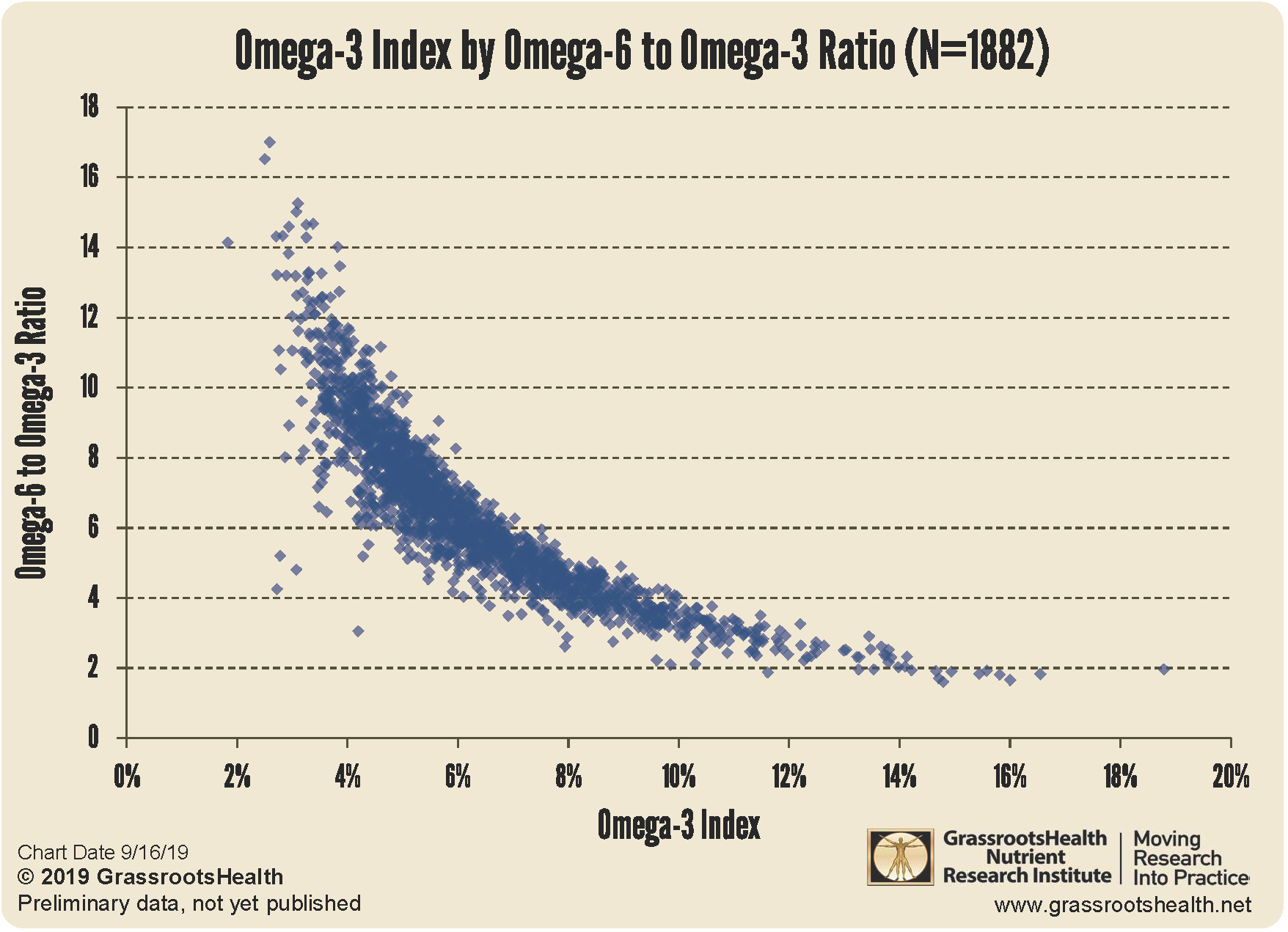
I quite like it as a measure because it is pretty damn difficult to get a lot of omega 3 in the red blood cells without having a low omega ratio.

So this is an interesting look at the potential results high vegetable oil, low fish vs low vegetable oil, high fish. Despite the usual epidemiological issues like healthy user bias.

It also identified stronger results with apoe4.

"Borderline statistical significance for a lower risk of AD was observed per standard deviation increase in RBC DHA (HR: 0.71, 95% CI: 0.51, 1.00, p = 0.053) in APOE-ε4 carriers"


Omega-3-to-6-by-Index-chart.png


Edited by aza, 17 January 2025 - 12:12 PM.

  • like x 1

sponsored ad

  • Advert
Click HERE to rent this advertising spot for BRAIN HEALTH to support LongeCity (this will replace the google ad above).

#8 aza

  • Topic Starter
  • Guest
  • 136 posts
  • 29
  • Location:aus
  • NO

Posted 18 January 2025 - 09:47 AM

glutathione, b vitamins and tau hyperphosphorylation

Its also worth noting that hydrogen sulphide should also be largely restored by glynac. This is important because HS may play a large role in preventing hyperphosphorylation of tau. B vitamins also appear to help here, making this an excellent combo to prevent progression.

“We further show that CSE is depleted in 3xTg-AD mice as well as in human AD brains, and that H2S prevents hyperphosphorylation of Tau by sulfhydrating its kinase, glycogen synthase kinase 3β (GSK3β). Finally, we demonstrate that sulfhydration is diminished in AD, while administering the H2S donor sodium GYY4137 (NaGYY) to 3xTg-AD mice ameliorates motor and cognitive deficits in AD.”

https://pubmed.ncbi....h.gov/33431651/


“8-month-old rats were injectedwith homocysteine via the vena caudalis with or without a concurrent folate/vit-B12 supplementation for 28 weeks. We found that hyperhomocysteinemia induced tau hyperphosphorylation and accumulation in hippocampus and cortex.”

“Folate/vit-B12 supplementation attenuated these bio-chemical and behavioural correlates.”

https://researchers....Wei_JAD2012.pdf


Homocysteine and autophagy

B vitamins unsurprisingly correct homocysteine induced autophagy downregulation (although some people might need activated b-vitamins). While glynac largely restores mitophagy and autophagy, it is possible that homocysteine lowering may just go the extra mile.


“Inhibition of autophagy by HHcy exacerbated cellular injury during oxygen and glucose deprivation and reperfusion (OGD/R), and oxidative stress. These effects were prevented by Vitamin B co-treatment, suggesting that it may be helpful in relieving detrimental effects of HHcy in ischemia/reperfusion or oxidative stress. Collectively, these findings show that Vitamin B therapy can reverse defects in cellular autophagy and ER stress due to HHcy; and thus may be a potential treatment to reduce ischemic damage caused by stroke in patients with HHcy.”

https://pmc.ncbi.nlm...les/PMC5260994/


Interestingly omega 3’s have an additive effect to reducing homo-cysteine. This may be one of the reasons why omega 3 and b vitamins appear to be much more effective together.


“vitamin B-12+fish oil supplementation lowered plasma Hcy concentrations by 22%, 19%, and 39%, respectively. “

https://pubmed.ncbi....h.gov/26420180/


Omega 3 + b vitamins just might be the kick in the pants glynac needs to almost fully restore mitophagy and autophagy to youthful levels. Among other things.


Edited by aza, 18 January 2025 - 09:49 AM.

  • Informative x 1


Also tagged with one or more of these keywords: alzheimers, glutathione, tau, amaloid

8 user(s) are reading this topic

0 members, 8 guests, 0 anonymous users