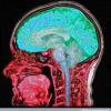
I have major problems with dopamine efficiency in the frontal cortex due to probable cell and efferent loss of dopamine from the left Caudate nucleus to the different areas of the frontal cortex.
Based on what Futurist 1000 has wrote,I have experienced these difficulties in the last 2 years........
Orbital syndrome :Emotional lability,Poor judgement and Insight,Distractability
Frontal convexity syndrome: Apathy,Psychomotor retardation,poor word list generation,Segmented visual-spatial analysis
Medial frontal syndrom(akinetic): Sparse verbal output,Lower extremity weakness,Gait abnormalities(frontal-cerebellum)ataxia>minor and bladder(urgency and frequency.
It appears that I have an ovoid lesion(8mmx3mm)from the blooming effect in the anterior portion of the left Head of the caudate nucleus,with a more mild susseptabilty of the globus pallidi(iron or mild calcification).
The origin remains a mystery.I have with the advice DDX of the original neuroradiologists ruled out Parkinson's,Huntington's,Wilsons disease,MS, Hallovordadn Shpatz variant,Hemachromatosis and Fahr's disease.
I dont have a history of hypertension and my MRA and CTA(CT angiogram) were negative for any large vessel disease or malformations.Although it's ability to view the anterior lenticulostriate arteries are limited.An MR with 7 tesla strength with a black blood aquasition sequence would be needed and is not available in North America yet for clinical purposes to my knowledge.However it is being used in Notingham England,where the MR first originated.
My MRI in Jan/07 shows this lesion to be very subtle compared to Mar/08 which the radiologists say has increased significantly based on T2 weighted and DWI sequences,howver no T2*Gradient echo was done in 2007.It has for the last 5 MRI's since Mar/08
I have had 2minor and 1 moderate concussions.1990,91 and 99 respectively, and my neurologist was insisting that this lesion was from the concussions.This from my knowledge was not possible as the forces were not severe enough to cause a shear injury.She sent me to a very intelligent Neurometabolic doctor who specializes in these non specific findings.
The junior Dr-assistant told me he thought it was a small stroke.The main doctor viewed my scans and felt that it was not a garden variety stroke and wanted to run a gambit of tests for Mitochondria disease.Blood tests MRI/MRS(Spectroscopy) and muscle biopsy.He is one of the best and deals with MIto diseases like Melas,MERFF,KKS which are genetic but can be brought on by certain neurotoxins.He also has expertise in CFS/ME and Fibromyalgia. If a Mito condition exists it can explain the propensity for a stroke-like lesion.
In 2007 I started noticing subtle cognitive problems but never gave much thought to it,but in Jan/08 I made a mistake and went to a rapid detox centre in USA for a CT withdrawal from a 14 year xanax addiction.I started on 0.5mgs and over the years it escalated to 4mgs,fighting all the time not to increase my dose.It was basically a cold turkey detox with bandaids-Neurontin,phenobarb and clonodine for Withdrawals! as needed?! Nothing first line for blood pressure which the records now show was 158/91 on discharge! I had some real bad palpitations the first 2 weeks and was not instructed to monitor my BP and the psych doctor in my province(Ontario) who agreed to monitor me post detox was ignorant and never inquired or instructed.
The few times I checked in Jan,Feb and March 04 they were within normal,but what happened for 2 weeks before BP was taken in Jan or after discontinuation of clonodine which they did NOT tell me to taper gradualy! I may have suffered from clonodine rebound hypertension.After my MRI on Mar.17/08 revealed this lesion which the first radiologist missed I was devestated and thought for sure I was damaged.
I wouldn't make it cold turkey anyway.
Sorry for all the details but I felt it was needed to give you an idea as to the complexity of the situation so you would be better suited to give me some possible advice.
Since 2008 I have symptoms of fatigue,abulia,Anxiety,processing and concentration problems.Retrieval difficulties and short/long term memory problems that were not there before.As well as some vocal tics.
I haven't been to the gym or sociable since this occured.
I was using pharm grade piracetam from a compounder(2400mgs/day) cobined with AOR citicholine and loved it.This was from 2005 to the end of 2007.I am more sensitive to all supplements now and am not sure that my symptom emergence and "lesion increase" are not from a failed withdrawal syndrome that might have caused a huge glutamate influx.
I am determined to try again slowly if I get approval from this Neurometabolic doctor.
I was considering Neuroreplete which was developed by Dr.Marty Hinz and has been shown to be clinically efective on neurotransmitter balance and repair instead of depletion from long term AD and stimulant medication.I was also going to restart my Pir/CDP choline combo.I have some Nootropil but it is expensive.What brand of bulk powder do you recommend?
I'd really appreciate any and all advice as to your thoughts of how to balance and increase dopamine afferent to the wounded caudate so that I can increase my efferent projections to the prefrontal cortex and mitigate my symptoms. I dont know how much can be improved but feel free to suggest any supps or meds that you would recomend and please look into Neuroreplete and tell me what you think.
Just an update> the neurometabolic doctors have found a slightly mild IgG cardiolipin level last week : 17 and it should be <15 although negative for antibodies is <6.
The hematologist did an IgM cardiolipin and a Lupus coagulant antibody to rule out phospholipd antibody syndrome.He doubts I have it,however even a mild positive result in IgG means that the antibodies are attacking my vessel walls and making me sensitive to clots(thrombosis) especially in the small feeding arteries and arterioles in subcortical structures.
BTW once 7Tesla MRI's are available in a clinical setting you will be able to visualize neuochemical activity instead of just structure because they will measure and track sodium changes and spin dynamics unlike regular MRI's which can only track proton molecules. I just had a SPECT on thursday.It probably wont add much but we'll see if there is any perfusion problems.
Left Head of Caudate via T2 Fast-Spin Echo Axial sequence

Left Head of Caudate on Coronal Gradient Echo

Left Head of Caudate via T2*Gradient Echo Axial sequence

This is less evident then the coronal gradient echo because of slice thickness 5mm vs.2mm
Left Head of caudate via Susceptability Weighted Image Axial sequence

Lesion is more evident and pronounced due to the susceptability of 3-D GRE and the BOLD component which is highly sensitive to signal loss from Iron or Hemosiderin
Other than Nootropics and medications to help with the prefrontal loss of dopamine input,I have to remain hopeful for the successful use of stem cell therapy to help me one day,as they have been used for Basal Ganglia strokes or other conditions like cerebral microbleeds and hopefully degenerative diseases like Huntingtons and Parkinsons.
Here is a link to a 60yr old lady who suffered Dysarthia from a Right-sided Caudate microbleed with a history of Hypertension.She presented in the acute phase with dysarthia and her lesion appears Hyperintense(bright white) on T1 and T2 Images because MRI is based on the deoxyhemoglobin in immediate scanning.Itis not until >14 days that the lesion will break down to hemosiderin that it can be seen as a low intensity T2 shortening(black)lesion on the gradient echo sequences accompanied by other smaller pettechial hemorhagic lesions on subsequent slices.
Caudate Microbleed
Thank you in advance to all advice,information and suggestions
Attached Files
Edited by NG_F, 12 September 2009 - 11:01 PM.


