• Log in with Facebook Log in with Twitter Log In with Google      Sign In    
  • Create Account
  LongeCity
              Advocacy & Research for Unlimited Lifespans

Photo
* * * * - 4 votes

Tickle Your Damn Amygdala's You Neurotic Fools!

amygdala visualization frontal lobes brain exercise

  • Please log in to reply
344 replies to this topic

#151 Mr Serendipity

  • Topic Starter
  • Guest
  • 986 posts
  • 19
  • Location:UK
  • NO

Posted 10 February 2020 - 08:40 PM

https://www.scienced...51026171805.htm


'Love hormone' helps produce 'bliss molecules' to boost pleasure of social interactions Role of oxytocin in triggering marijuana-like neurotransmitters uncovered Date: October 26, 2015 Source: University of California - Irvine Summary: The hormone oxytocin, which has been associated with interpersonal bonding, may enhance the pleasure of social interactions by stimulating production of marijuana-like neurotransmitters in the brain, according to a new study. The research provides the first link between oxytocin -- dubbed the 'love hormone' -- and anandamide, which has been called the 'bliss molecule' for its role in activating cannabinoid receptors in brain cells to heighten motivation and happiness.
FULL STORY
 

The hormone oxytocin, which has been associated with interpersonal bonding, may enhance the pleasure of social interactions by stimulating production of marijuana-like neurotransmitters in the brain, according to a University of California, Irvine study.

The research provides the first link between oxytocin -- dubbed the "love hormone" -- and anandamide, which has been called the "bliss molecule" for its role in activating cannabinoid receptors in brain cells to heighten motivation and happiness. Results appear the week of Oct. 26 in the early online edition of Proceedings of the National Academy of Sciences.

To investigate the role of anandamide in social contact, UCI's Daniele Piomelli -- the Louise Turner Arnold Chair in the Neurosciences and founding director of the drug discovery & development department at the Italian Institute of Technology in Genoa, Italy -- and his colleagues measured levels of this marijuana-like neurotransmitter in mice that had been either isolated or allowed to interact. Anandamide is among a class of naturally occurring chemicals in the body known as endocannabinoids that attach to the same brain cell receptors as does marijuana's active ingredient, THC, with similar outcomes.

The researchers discovered that social contact increased production of anandamide in a brain structure called the nucleus accumbens, which triggered cannabinoid receptors there to reinforce the pleasure of socialization. When cannabinoid receptors were blocked, this reinforcement disappeared.

Piomelli's team then looked for a possible connection between anandamide and oxytocin, which is well known for its role in promoting social contact. A small number of neurons in the brain make oxytocin and use it as a neurotransmitter. When the scientists stimulated those neurons, they saw an increase in anandamide creation in the nucleus accumbens. More importantly, they found that blocking anandamide's effects also blocked the pro-social effects of oxytocin, which implies that oxytocin reinforces social ties by inducing anandamide formation.

Adding medical interest to this discovery, the researchers showed that interrupting anandamide degradation enhanced the pleasure of social contact. Animals treated with a drug that stops anandamide degradation behaved as though they enjoyed spending time with their cage mates more than animals treated with a placebo, Piomelli noted.

Oxytocin has also been called the hug hormone, cuddle chemical and moral molecule due to its effects on behavior, including its role in love and female reproductive functions. A 2011 study by Dutch scientists revealed that oxytocin makes people feel more extroverted, and clinical researchers are investigating it as a possible treatment for the symptoms of autism. But it's very hard to deliver oxytocin, a small protein, to the human brain.

"Our findings open the exciting possibility that drugs that block the degradation of anandamide, which are currently being tested for various anxiety disorders, could give a boost to the brain's own oxytocin and help people with autism socialize more," Piomelli said.


#152 Mr Serendipity

  • Topic Starter
  • Guest
  • 986 posts
  • 19
  • Location:UK
  • NO

Posted 10 February 2020 - 08:49 PM

https://www.ncbi.nlm...pubmed/16224541


Cannabinoids promote embryonic and adult hippocampus neurogenesis and produce anxiolytic- and antidepressant-like effects.
Author information
Abstract

The hippocampal dentate gyrus in the adult mammalian brain contains neural stem/progenitor cells (NS/PCs) capable of generating new neurons, i.e., neurogenesis. Most drugs of abuse examined to date decrease adult hippocampal neurogenesis, but the effects of cannabis (marijuana or cannabinoids) on hippocampal neurogenesis remain unknown. This study aimed at investigating the potential regulatory capacity of the potent synthetic cannabinoid HU210 on hippocampal neurogenesis and its possible correlation with behavioral change. We show that both embryonic and adult rat hippocampal NS/PCs are immunoreactive for CB1 cannabinoid receptors, indicating that cannabinoids could act on CB1 receptors to regulate neurogenesis. This hypothesis is supported by further findings that HU210 promotes proliferation, but not differentiation, of cultured embryonic hippocampal NS/PCs likely via a sequential activation of CB1 receptors, G(i/o) proteins, and ERK signaling. Chronic, but not acute, HU210 treatment promoted neurogenesis in the hippocampal dentate gyrus of adult rats and exerted anxiolytic- and antidepressant-like effects. X-irradiation of the hippocampus blocked both the neurogenic and behavioral effects of chronic HU210 treatment, suggesting that chronic HU210 treatment produces anxiolytic- and antidepressant-like effects likely via promotion of hippocampal neurogenesis.


sponsored ad

  • Advert
Advertisements help to support the work of this non-profit organisation. To go ad-free join as a Member.

#153 Mr Serendipity

  • Topic Starter
  • Guest
  • 986 posts
  • 19
  • Location:UK
  • NO

Posted 10 February 2020 - 08:54 PM

AND NOW FOR THE MOST IMPORTANT AND RELEVANT STUDY ON ANANDAMIDE AND THE AMYGDALA & FRONTAL LOBES!


https://www.nature.com/articles/ncomms7395


FAAH genetic variation enhances fronto-amygdala function in mouse and human

Nature Communications volume 6, Article number: 6395 (2015Cite this article

Abstract

Cross-species studies enable rapid translational discovery and produce the broadest impact when both mechanism and phenotype are consistent across organisms. We developed a knock-in mouse that biologically recapitulates a common human mutation in the gene for fatty acid amide hydrolase (FAAH) (C385A; rs324420), the primary catabolic enzyme for the endocannabinoid anandamide. This common polymorphism impacts the expression and activity of FAAH, thereby increasing anandamide levels. Here, we show that the genetic knock-in mouse and human variant allele carriers exhibit parallel alterations in biochemisty, neurocircuitry and behaviour. Specifically, there is reduced FAAH expression associated with the variant allele that selectively enhances fronto-amygdala connectivity and fear extinction learning, and decreases anxiety-like behaviours. These results suggest a gain of function in fear regulation and may indicate for whom and for what anxiety symptoms FAAH inhibitors or exposure-based therapies will be most efficacious, bridging an important translational gap between the mouse and human.

Introduction

Translational research holds the promise to leverage basic scientific findings into advances for human health. Studies in animal models play an important role in this process, enabling the precise delineation of the mechanisms underlying behaviour in humans, in whom such fine-scale resolution is difficult to achieve. A critical requirement for the success of this translational approach is that the phenotypes of interest are consistent across species. In the present study, we show that a single-nucleotide polymorphism (SNP) in the fatty acid amide hydrolase (FAAH) gene of the endocannabinoid system has parallel molecular, neural and behavioural effects in both humans and mice engineered to express the variant human allele, filling an important translational gap.

The endocannabinoid system has been implicated in human anxiety1,2,3. Anandamide (AEA), an endogenous cannabinoid (eCB) agonist at the CB1receptor, is proposed to play a central role in this effect2,4. FAAH is a catabolic enzyme and primary regulator of AEA signalling in the brain5. In humans, differential expression of FAAH protein is associated with a common SNP (C385A; rs324420) of which ~38% of individuals of European descent are carriers (AC and AA genotypes)6. This polymorphism leads to the substitution of an evolutionarily conserved proline at amino-acid position 129 with a threonine residue, which in turn renders the FAAH protein more vulnerable to proteolytic degradation. Accordingly, the FAAH C385A SNP enhances eCB signalling by reducing steady-state levels of FAAH protein, which leads to elevated AEA levels7,8,9. Pharmacologic manipulations and genetic knockout of FAAH have been implicated in anxiolytic behaviours including enhanced fear extinction learning2,10,11. However, the ability to characterize the effects of the FAAH variant in the brain has been limited, since the variant is only present in humans. Here, we describe the development of a knock-in mouse that expresses the variant A (threonine) allele of the FAAH polymorphism in place of the conserved ancestral C (proline) allele, enabling the demonstration of parallel molecular, circuit-level and behavioural phenotypes in humans and in the knock-in mice carrying this variant.

Results
Generation and validation of FAAH C385A knock-in mice

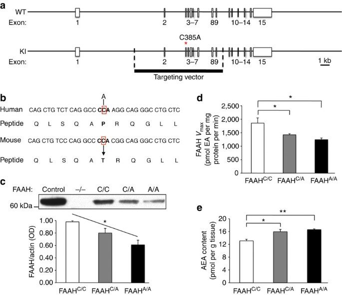
We examined molecular and biochemical effects in the FAAH knock-in mouse to determine whether they paralleled effects of the human SNP (Fig. 1a,b). Specifically, human carriers of the A-allele have been shown to have reduced FAAH protein levels in their lymphocytes due to protein-folding abnormalities and increased proteolytic breakdown, leading to elevated plasma levels of AEA7,8,9. A fundamental question has been whether these alterations observed in peripheral tissues reflect parallel modulation of the eCB system by this SNP in the brain. In the FAAH knock-in mouse, analysis of relative protein expression levels in the forebrain showed a main effect of genotype on FAAH protein levels (analysis of variance (ANOVA) with post hoc Dunnett’s test (F(2,6)=8.96, P<0.02)) with an allele dose-dependent decrease in FAAH levels among knock-in mutants (Fig. 1cSupplementary Fig. 1). There was also a main effect of genotype on FAAH hydrolytic activity (ANOVA with post hoc Dunnett’s test (F(2,12)=7.89 P<0.01)) (Fig. 1d) and AEA levels (ANOVA with post hoc Dunnett’s test (F(2,11)=7.25, P<0.02)) (Fig. 1e), but not for the levels of the endocannabinoid, 2-arachidonoylglycerol (2-AG), which is not a FAAH substrate (Supplementary Fig. 2). There was also no genotypic effect on the maximal binding site density for the CB1receptor (Supplementary Fig. 3). This mouse model provides the first demonstration of biochemical changes within the brain due to the FAAH C385A SNP and mirrors its reported effects in human lymphocytes. These findings validate that the FAAH C385A knock-in mouse recapitulates the known molecular and biochemical phenotypes of the human FAAH C385A polymorphism, supporting its use as a model of higher-level neural and behavioural phenotypes.

Figure 1: Generation and validation of FAAH C385A knock-in mice.
41467_2015_Article_BFncomms7395_Fig1_HTM

(a) A portion of the coding region in the FAAH gene is replaced with C385A SNP. (b) The region encompassing the SNP has high homology between human and mouse FAAH genes. © FAAH protein levels in knock-in mice (FAAHC/A; FAAHA/A) and wild-type (FAAHC/C) littermates from three independent western blot analyses. Brain homogenates from FAAH−/− mice, and lysates from heterologous 293 cells overexpressing FAAH, were used as controls. (d) FAAH hydrolytic activity and (e) anandamide (AEA) content in FAAH knock-in mouse forebrain homogenates (n=4 per group). (EA; ethanolamine). Means±s.e.m. presented. *P<0.05, **P<0.01.

Enhanced fronto-amygdala connectivity in humans and mice

We tested for cross-species translation in genotypic effects of FAAH on fronto-amygdala circuitry and function in mice and humans with the variant A-allele. In humans, FAAH C385A has been associated with variation in reactivity to threat1. However, it is unclear how the FAAHC385A polymorphism might alter the circuitry implicated in this behavioural domain. Fear-conditioning studies in animal models suggest that dynamic interaction between the amygdala and two subregions of the prefrontal cortex (PFC) can drive opposing behavioural responses to threat12,13,14,15. Whereas the prelimbic (PL) region promotes fear expression, the infralimbic (IL) region constrains the regulation of threat responses. Neuroimaging studies examining correlates of these opposing behaviours in humans suggest that a dorsal anterior cingulate cortex (ACC) region exhibits functional parallels to rodent PL, and a subgenual region of ventromedial PFC (vmPFC) exhibits functional parallels to rodent IL16,17,18,19.

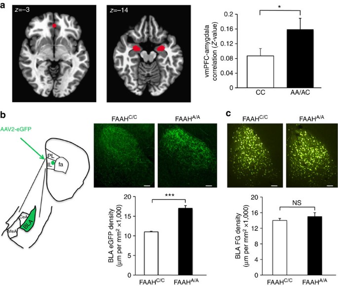
We performed species–specific connectivity analyses focusing on key regions within the fronto-amygdala circuitry that regulate fear responses and fear extinction learning. First, we examined resting-state connectivity between the subgenual vmPFC and amygdala and the dorsal ACC and amygdala in humans by genotype. A-allele carriers (Supplementary Table 1) showed increased correlations between the blood oxygen level-dependent signals in the vmPFC and the bilateral amygdala (t(34)=2.71, P=0.037) (Fig. 2aSupplementary Fig. 4a; Bayesian statistical analysis in Supplementary Fig. 5a). This pattern was selective to subgenual vmPFC–amygdala connectivity, with no genotypic difference in dorsal ACC–amygdala connectivity (Supplementary Fig. 6a). A substantial literature suggests that stronger inverse functional connectivity between the vmPFC and the amygdala during emotion-related tasks is associated with decreased anxiety or negative emotion20,21,22. In contrast, positive resting-state functional connectivity between these regions has been associated with lower anxiety23,24,25. Our present finding is consistent with this broader literature suggesting that greater positive corticoamygdala connectivity at rest predicts more effective emotional control23,26.

Figure 2: Functional and structural connectivity between ventromedial prefrontal cortex (vmPFC) and amygdala in humans and mice with FAAH C385A.
41467_2015_Article_BFncomms7395_Fig2_HTM

(a) fMRI functional connectivity compared between subgenual vmPFC (x, y, z=0, 40, −3) and bilateral amygdala in A-allele carriers (n=17) relative to C homozygotes (n=18). (b) Anterograde tracer (AAV2-eGFP; eGFP), targeted to IL, labelled afferents in BLA in FAAHA/A mice (n=4) and controls (FAAHC/Cn=4). Drawing illustrates anatomical boundaries. © Retrograde tracer (fluorogold; FG), targeted to IL, labelled BLA cell bodies in FAAHA/A mice (n=4) and controls (FAAHC/Cn=4). (Scale bars, 100 μm.) Means±s.e.m. presented. *P<0.05, ***P<0.001. NS, not significant.

To delineate the precise location and directionality of genotypic differences in fronto-amygdala circuitry, we used anterograde (adeno-associated virus 2 expressing enhanced green fluorescent protein; AAV2-eGFP) and retrograde (fluorogold (FG)) tracers in our FAAH knock-in mouse. These tract-tracing experiments revealed increased projections from IL to basolateral amygdala (BLA) in mice (FAAHA/A>FAAHC/C; two-tailed Student’s t-test, P<0.001), but no genotypic differences in ascending projections from BLA to IL (FAAHA/A>FAAHC/C; two-tailed Student’s t-test, P>0.56) (Fig. 2b,c). Mirroring the human imaging findings, we found no genotypic differences in descending or ascending projections between the PL and BLA in the mouse (Supplementary Fig. 6a–c). Selective increases in descending IL-amygdala projections offer a structural neuroanatomical basis for the increased functional connectivity in fronto-amygdala circuitry in human A-allele carriers and may help explain reported genotypic differences in emotion regulation1.

Enhanced cued fear extinction in humans and mice

Building on our biochemical and circuit-level genotypic differences in mice and humans, we tested for behavioural effects of this variation across species in parallel fear extinction experiments. The selective increase in connectivity within an established fear regulatory circuit (IL–BLA projection) in the mouse and the homologous vmPFC–amygdala circuit in human, led to the a priori prediction that A-allele carriers would show improved fear extinction learning but no difference in fear acquisition. These predictions are consistent with reported effects of pharmacological and genetic knockout manipulations of FAAH expression in mice2,27,28. We tested humans (22 CC-alleles, 18A-allele carriers; Supplementary Table 2) and mice using cued fear conditioning with extinction on the following day. In humans, there was no main effect of the FAAH genotype on fear acquisition (Supplementary Fig. 7a), but there was an effect of the genotype on fear extinction. Namely, human A-allele carriers exhibited facilitated fear extinction learning, as indexed by decreased galvanic skin responses during late trials of extinction training (F(1,36)=4.35, P=0.044), controlling for age and gender effects between genotypic groups (Fig. 3aSupplementary Fig. 4b; Bayesian statistical analysis in Supplementary Fig. 5b). Parallel findings in mice showed a significant effect of genotype on freezing behaviour during extinction (ANOVA with post hoc Bonferroni test (F(2,116)=6.8, P<0.01)) (Fig. 3b), with heterozygous (P<0.05) and homozygous (P<0.01) FAAH C385A mice showing less freezing behaviour compared with wild types in late trials, but no difference in early trials (Fig. 3b) or in fear acquisition (Supplementary Fig. 7b). These parallel effects of enhanced fear extinction in FAAH A-allele carriers have not previously been established for both mice and humans.

Figure 3: Enhanced cued fear extinction in humans and mice with FAAH C385A.
41467_2015_Article_BFncomms7395_Fig3_HTM

(a) Extinction, indexed by differential skin conductance response (SCR) (CS+–CS−), in 18 A-allele carriers and 22 C homozygotes. Trials were binned into early (average of the first five trials) and late (average of the following six trials). (b) Fear extinction, time spent freezing to cue, was tested in wild-type (FAAHC/Cn=21), heterozygous (FAAHC/An=20) and homozygous knock-in mice (FAAHA/An=20). Extinction trials were binned into early (average of extinction day 1) and late trials (average of extinction day 4). Means±s.e.m. presented. *P<0.05, #P<0.05 heterozygous knock-in mice versus wild-type controls, **P<0.01 homozygous knock-in mice versus wild-type controls.

Decreased anxiety levels in humans and mice

We further characterized the anxiolytic phenotype of the FAAH variant A-allele in both mice and humans using standard measures of anxiety-related behaviours. Given that our results demonstrated enhanced fear extinction in human and mouse FAAH A-allele carriers, we predicted they would also show reduced anxiety1,2,10,27,28,29. In 137 humans (Supplementary Table 3), A-allele carriers reported reduced levels of trait anxiety (t(135)=2.30, P=0.019) (Fig. 4aSupplementary Fig. 4c; Bayesian statistical analysis in Supplementary Fig. 5c); this effect was replicated in our separate fear extinction cohort (Supplementary Table 2Supplementary Fig. 8). In mice, we performed two standard measures of anxiety-like behaviours that involve placing subjects in conflict situations. In the elevated plus maze (EPM) test, homozygous mutant mice spent a higher percentage of time in the open arms than wild-type controls (ANOVA with post hoc Dunnett’s test (F(2,37)=2.89, P<0.04)), indicative of reduced anxiety-like behaviour (Fig. 4b). There was no significant difference in total distance travelled (Supplementary Fig. 9). In the novelty-induced hypophagia (NIH) task, mice were trained to approach a reward (sweetened milk) in their home cage and then tested in a novel, brightly lit environment. For this task, the latency to approach and drink the sweetened milk in the novel environment is a measure of anxiety-related behaviour30. FAAH C385A mice showed decreased latency to drink milk in the NIH task, suggesting decreased anxiety phenotype (ANOVA with post hoc Dunnett’s test (F(2,44)=5.83, P<0.01)) (Fig. 4c). To investigate the neural correlates of the observed anxiolytic effects, we examined neural activity, as indicated by expression of the early immediate gene c-Fos31, in the BLA following the NIH task. We found a main effect of genotype on BLA c-Fos expression in mutant knock-in mice (ANOVA with post hoc Dunnett’s test (F(2,15)=53.92, P<0.0001)) in a dose-dependent pattern, suggesting decreased engagement of the BLA in these mice during a stressful experience (Fig. 4d). This reduced activation of the amygdala in response to stressful situations is consistent with reductions in amygdala activity in response to threat in human FAAH A-allele carriers1.

Figure 4: Decreased anxiety in humans and mice with FAAH C385A.
41467_2015_Article_BFncomms7395_Fig4_HTM

(a) Trait anxiety levels in A-allele carriers (n=57) and C homozygotes (n=80). Anxiety-related behaviour in FAAH C385A mice in (b) elevated plus maze (FAAHC/Cn=17; FAAHC/An=11; FAAHA/An=12) and © novelty-induced hypophagia (NIH) (FAAHC/Cn=16; FAAHC/An=21; FAAHA/An=10). (d) Photomicrographs of NIH-induced c-Fos (red) labelling in the BLA (parvalbumin (green)) in FAAH C385A mice. Density of c-Fos immunoreactive (IR) cells in BLA, n=5 per group. (Scale bars. 50 μm upper row, 100 μm lower row.) Means±s.e.m. presented. *P<0.05, **P<0.01, ****P<0.0001.

Discussion

Using a vertically integrated approach with parallel studies in humans and mice, we have identified the relevance and impact of the FAAHC385A polymorphism on brain biochemistry, neurocircuitry, behaviour and symptoms. We validate our mouse model by showing that the FAAHvariant allele leads to reductions in FAAH protein and enzymatic activity and increases in AEA levels in the brain. Our subsequent analyses in humans and mice elucidated circuit level and behavioural phenotypes not previously established in human carriers of this variant. We found enhanced fear extinction in both humans and mice with the C385A mutation. In addition, our finding of selectively enhanced fronto-amygdala connectivity in both mouse and human A-allele carriers provides a mechanistic explanation for these behavioural effects through enhanced regulation of BLA responses to threat by the IL. Non-replication of candidate gene association studies has been a major problem within the field of behavioural genetics. Here, we have used a parallel mouse–human experimental approach that enables greater control for environmental and genetic confounds. The convergent findings reported here establish that effects of genetic variation in FAAH are evident at neural and behavioural levels in both the mouse and in humans, providing a persuasive cross-species validation. Thus, this variant SNP may represent a gain of function in the domain of anxiety-related behaviours and may prove valuable in determining for whom and for what symptoms FAAH inhibitors or exposure-based therapies, which build on basic principles of extinction learning, will be most efficacious. In this way, therapeutics might be tailored to an individual to move from standard care to more precise personalized care. A persistent problem identified in animal models of disease is a failure of the findings to translate to the human in clinical trials. Our studies translate mouse behavioural and brain findings to show their human relevance using parallel paradigms and imaging tools across species. Thus, the present results obtained using this integrated approach bridge a large translational gap from mouse to human.

Methods
Generation of FAAH C385A mice

The replacement-targeting vector consisted of a 9.53-kb mouse genomic fragment containing exons 2–9 FAAH-coding sequence. In a 1.6-kb fragment containing the FAAH-coding region, the C385A mutation was introduced and put back into the targeting vector. The flippase recognition target Neo cassette was introduced via homologous recombination as a positive selection marker. A pGK-thymidine kinase cassette was used as a negative selection marker. The targeting vector was comprised of a 1.88-kb short arm, a 5.52-kb long arm, a 9.53-kb targeted sequence carrying the C385A mutation and the Neo cassette flanked by two flippase recognition target sites. Linearalized targeting vectors were electroporated into iTL BA1 (C57BL/6N × 129/SvEv) hybrid embryonic stem cells. DNA derived by G418-resistant embyonic stem cell clones were screened using a diagnostic SspI and BamHI restriction enzyme digestions. Recombinant clones containing the predicted 8.6- and 10.4-kb rearranged bands were obtained at a frequency of 5 in 100. Two positive embryonic stem cell clones were injected into C57BL/6 blastocysts, which were then introduced into pseudopregnant females. Chimeric animals were mated with C57BL/6 to produce heterozygous animals, and these mice were subsequently crossed with C57LB/6 mice expressing FLP recombinase in germ cells to excise the neo cassette. Upon weaning, all genotypes were housed together and randomly assigned to experimental groups. Sample size was chosen based on previous literature for each experimental procedure. All analyses were performed on mice back-crossed at least seven generations onto the C57BL/6 genetic background.

Western Blot analysis

Samples of brain lysates were prepared as described previously32. Briefly, brain tissue was incubated in RIPA lysis buffer (150 mM NaCl, 50 mM Tris pH 8, 5 mM EDTA, 1% Triton X-100, 0.5% sodium deoxycholate and 0.1% sodium dodecyl sulphate) with protease and phosphatase inhibitors (1:100, Calbiochem #539131 and #524625, respectively). Samples were triturated, rotated at 4 °C for 30 min then spun down at 14,000 r.p.m. for 10 min at 4 °C. The supernatant was collected and cell debris discarded. Protein concentration was measured using a BCA kit. To measure FAAH levels, 10 mg of forebrain lysate was resolved by SDS–PAGE electrophoresis (NuPAGE 10% Bis-Tris gel; Invitrogen #NP0315) and probed with antibodies: mouse monoclonal anti-FAAH (Abcam, ab54615; 1:500) and goat polyclonal anti-actin (Santa Cruz, sc-1616HRP; 1:5,000) were used. As a positive control, 293T cells purchased from American Type Culture Collection were transfected with 1 mg mouse FAAH complementaryDNA clone (Origene, NM_010173), and lysates were processed in parallel. Forebrain lysate from FAAH knockout mice5were used as a negative control.

FAAH activity assay

FAAH activity from forebrain lysates was measured as the conversion of AEA labelled with [3H] in the ethanolamine portion of the molecule ([3H]AEA) to [3H]ethanolamine, as reported previously33. Membranes were incubated in a final volume of 0.5 ml of TME buffer (50 mM Tris-HCl, 3.0 mM MgCl2 and 1.0 mM EDTA, pH 7.4) containing 1.0 mg ml−1fatty acid-free bovine serum albumin (BSA) and 0.2 nM [3H]AEA. Isotherms were constructed using eight concentrations of AEA at concentrations between 10 nM and 10 μM. Incubations were carried out at 37 °C and were stopped with the addition of 2 ml of chloroform/methanol (1:2). After standing at ambient temperature for 30 min, 0.67 ml of chloroform and 0.6 ml of water were added. Aqueous and organic phases were separated by centrifugation at 1,000 r.p.m. for 10 min. The amount of [3H] in 1 ml of the aqueous phase was determined by liquid scintillation counting and the conversion of [3H]AEA to [3H]ethanolamine was calculated. The Vmax values for this conversion were determined by fitting the data to a single-site competition equation using Prism.

Lipid extraction from tissue for eCB levels

For analysis of endocannabinoid content, brain regions were subjected to a lipid extraction process. Forebrain tissue samples were weighed and placed into borosilicate glass culture tubes containing 2 ml of acetonitrile with 5 pmol of [2H8]anandamide and 5 nmol of [2H8]2-arachidonoylglycerol for extraction. Tissue was homogenized with a glass rod and sonicated for 30 min on ice. Samples were incubated overnight at −20 °C to precipitate proteins then centrifuged at 1,500 gto remove particulates. The supernatants were removed to a new glass tube and evaporated to dryness under N2 gas. The samples were resuspended in 300 μl of acetonitrile to recapture any lipids adhering to the glass tube, and dried again under N2 gas. Finally, lipid extracts were suspended in 20 μl of acetonitrile, and stored at –80 °C until analysis.

Mass spectrometrical detection of eCBs

Liquid chromatography–mass spectrometry (LC–MS/MS) analyses were carried out on an Eksigent ekspert micro LC 200 coupled with an AB Sciex Qtrap 5500 mass spectrometer, installed with a Turbo V Spray ion source (AB Sciex, Canada). The LC was equipped with a temperature-controlled CTC autosampler. An Eksigent HALO C18 HPLC column (1 × 50 mm, 2.7 μm particle diameter, 90 Å pore size) was used. Samples were analysed isocratically, at a flow rate of 30 μl min−1, and a solvent composition of 15% mobile phase A (10 mM ammonium acetate in water) and 85% mobile phase B (acetonitrile). After 3.25 min, the column was regenerated with 100% B. Before each injection, the column was re-equilibrated at the initial mobile phase condition for 2 min. Following each LC–MS/MS run, a blank was run. The sample injection loop (5 μl loop size) was rinsed with 40 μl of methanol, and the column purged for 5 min with 100% B for 4.5 min and followed by 85% B for 0.5 min. This was intended to mitigate cross contaminations due to carryover from preceding sample injections. The LC column was maintained at 25 °C, and the samples at 10 °C.

The mass spectrometer was operated in positive ion mode, with the ion-spray voltage set at 5,500 V, curtain gas at 20 (a.u.), source gas 1 and gas 2 both at 40 (a.u.), and source temperature at 300 °C. Protonated molecular ions of AEA (m/z 348) and AEA-d8 (m/z 356), and ammonium adduct ions of 2-AG (m/z 396) and 2-AG-d8 (m/z 404) were selected as the respective precursor ions for collision induced dissociation (CID). Multiple reaction monitoring (MRM) scan modes were used with Q1 and Q3 both at unit resolution. Optimized collision energies for the transitions were as follows: AEA (348 to 62) CE 22 V, AEA-d8 (356 to 62) CE 22 V, 2-AG (396 to 287) CE 15 V and 2-AG-d8 (404 to 294) CE 17 V.

Each brain tissue extract sample was further diluted in acetonitrile to yield a 100-fold diluted sample, for the LC–MS/MS quantification of 2-AG, while the undiluted extract sample was analysed directly for quantification of AEA. It has been reported that 2-AG undergoes spontaneous isomerization converting to 1-AG by acyl group migration during tissue extraction and reconstitution procedures. We also observed that 1-AG was present in authentic standard solutions of 2-AG, as well as brain tissue extracts. Peak areas of 1-AG and 2-AG were combined to establish a standard calibration curve for 2-AG. The data processing was accomplished using Analyst 1.5.2 software (AB Sciex). Linear regressions of relative peak areas (analyte to internal standard ratios) were performed over analyte concentrations from 0.00025 to 0.25 pmol μl−1 (AEA), and 0.0025 to 2.5 pmol μl−1 (2-AG). Analyte levels were normalized to tissue weight.

CB1 receptor-binding assay

CB1 receptor agonist-binding parameters were determined using radioligand binding using a Multiscreen Filtration System with Durapore 1.2-lM filters in 96-well filter plates (Millipore, USA) through a protocol previously described34. Incubations (total volume=0.2 ml) were carried out using TME buffer containing 1 mg ml−1 BSA (TME/BSA). Membranes (10 μg protein per incubate) were added in triplicate to wells containing 0.1, 0.25, 0.5, 1.0, 1.5 or 2.5 nM [3H]CP 55,940 (American Radiochemicals, USA), a cannabinoid CB1 receptor agonist, and incubated for 1 h at room temperature on an orbital shaker. Ten micromolar AM251 (Tocris Biosciences, USA) was used to determine nonspecific binding. Bmax (maximal binding site density) values were determined by nonlinear curve fitting of specific binding data to the single site-binding equation using GraphPad Prism (GraphPad, USA).

Behavioural overview

To reduce experimental variability, age-matched littermate pairs resulting from heterozygous crossings were randomly assigned to experimental groups. Only adult male mice were used for all experiments. All behavioural measurements were performed by observers blind to genotype. All animals were kept on a 12:12 light–dark cycle at 18 °C~22 °C with food and water available ad libitum unless noted otherwise. All procedures were in accordance with the NIH Guide for the Care and Use of Laboratory Animals and were approved by the Institutional Animal Care and Use Committee of Weill Cornell Medical College.

Cued fear-conditioning and extinction procedure

The task was conducted as described previously35. Briefly, the fear-conditioning apparatus consisted of a mouse shock-chamber (Coulbourn Instruments, USA) placed in a sound-attenuated box. On day 1 (acquisition), after a 2-min acclimation period to the conditioning chamber (scented with 0.1% peppermint in 70% EtOH), mice were fear conditioned with three tone–shock pairings, consisting of a 30-s presentation of a tone (conditioned stimulus; 5 kHz, 70 dB) that coterminated with a 0.7-mA foot shock (unconditioned stimulus) during the last 1.0 s of the tone with an intertrial interval of 30 s. Mice remained in the conditioning chamber for 1 min before being returned to their home cages. Twenty-four hours after fear acquisition, mice were exposed to five presentations of the tone in the absence of shock (extinction). To eliminate any confounding interactions of contextual fear, tones were presented in a novel context, consisting of a white cylindrical arena (scented with 0.1% lemon in 70% EtOH). Tone presentations lasted for 30 s with an intertrial interval of 30 s. After the final tone presentation, mice remained in the conditioning chamber for 1 min before being returned to their home cages. Fear extinction trials were repeated daily for a total of 4 days of extinction training. Graphic State 3.02 software was used to control experiments. Mice were videotaped for subsequent analysis. Freezing was defined as the absence of visible movement except that required for respiration36. The percentage time spent freezing was calculated by dividing the amount of time spent freezing during the 30-s tone presentations by the duration of the tone.

EPM procedure

The EPM task was conducted as described previously37. The maze was constructed of grey Plexiglas, raised 70 cm above the floor and consisted of two opposite closed arms with 14-cm-high opaque walls and two opposite open arms of the same size (29 cm × 6 cm). The maze was set up under a digital camera connected to a video recorder and computer under the control of Ethovision XT 5.0 software (Noldus, USA) to live track subject movements. A single testing session of 10 min was carried out under dim light (~6 Lux in centre of maze). To begin a trial, the animal was placed in the centre of the maze facing an open arm. The maze was cleaned with a 70% ethanol solution between trials to eliminate possible odour cues left by previous subjects. Measures of anxiety-like behaviour were assessed by calculating the percentage time spent in the open arms by dividing the amount of time spent in the open arms by the duration of the trial (10 min).

NIH procedure

The task was conducted as described previously38. Briefly, mice received 3 consecutive days of training (days 1–3) prior to testing in a room with dim lighting to acclimate to the milk and the manner in which it is administered (metal nozzle). Training consisted of presenting the mice with a standard dual-bearing sipper tube (5 oz. bottle) inserted between the wire bars of the home cage lid containing 90% sweetened condensed milk diluted in tap water. On day 4, mice underwent home cage testing in which all mice were placed into a holding cage and then returned to the home cage one at a time for testing in dim lighting. The latency to drink was recorded over a 10-min period. Mice that did not approach the sipper bottle during the home cage test were excluded from the study (four mice in total). There were no baseline differences in latency to drink between groups in the home cage test. On day 5, novel cage testing was conducted by placing a single mouse into a clean cage with no bedding of the same dimensions as their home cage, but under bright conditions and with a white piece of paper under the cage. Excess light was focused specifically on the sipper bottle. Mice were again presented with a sipper tube containing 90% sweetened milk, and the latency to drink over a 10-min period was reported as a measure of anxiety-like behaviour.

c-Fos immunohistochemistry

All experiments were carried out at room temperature, as previously described38, unless otherwise specified. Ninety minutes after exposure to experimental factors, mice were sacrificed by intraperitoneal injection of Euthasol and perfused through the heart with 30 ml of saline followed by 90 ml of 4% paraformaldehyde in 0.1 M sodium phosphate (pH 7.4) at flow rate of 30 ml min−1. Brains were removed and post-fixed overnight in 4% paraformaldehyde before being transferred to 30% sucrose in 0.1 M sodium phosphate (pH 7.4) for 48 h at 4 °C. Brains were frozen in powdered dry ice and stored at −20 °C until sectioning. Coronal sections (40 μm) of whole brain were cut by using sliding microtome frozen by powdered dry ice. Six sets of serial sections were collected in Ependoff tubes each containing 2 ml cryoprotectant (30% glycerol and 30% ethylene glycol in 0.1 M sodium phosphate, pH 7.4) and stored at −20 °C. Free-floating serial sections (take one every third) were washed (three times for 10 min each) in TBS, incubated for 30 min in a blocking solution containing 4% normal horse serum and 1% BSA in Tris buffered saline (TBS) with 0.2% Triton X-100 (TBS–Tx) and incubated overnight at 4 °C with rabbit anti-c-Fos primary antibody (c-Fos sc-52, Lot# F2209, Santa Cruz Biotechnology, USA; sc-52 antibody was raised against amino acids 3–16 of human c-Fos: SGFNADYEASSSRC) diluted 1:1,000 mixed with goat anti-parvalbumin primary antibody (1:2,000, Lot# PVG 214, Swant, Switzerland) in the blocking solution mentioned above. Sections were then washed in TBS and incubated for 2 h with Alexa Fluor-labelled donkey anti-rabbit and anti-goat IgG secondary antibodies (Alexa Fluor 555 and 488) diluted 1:500 in TBS–Tx. Sections were again washed, mounted on chromalum/gelatin-coated slides and air-dried for 2 h in dark. Slides were cover-slipped by water-soluble glycerol-based mounting medium containing 4',6-diamidino-2-phenylindole (DAPI), and sealed with nail polish. Estimation of cell density of c-Fos-positive neurons in amygdala was performed with StereoInvestigator 9.0 (Microbrightfield, USA). Briefly, serial sections (every third section—120 μm were numbered by rostra-caudal order, and contours of BLA was traced by referring to the Allen Brain Map (Allen Co., USA). For the boundaries of the amygdala, we used parvalbumin counter-stain combined with DAPI to yield a clear boundary for subnuclei or layers. All cells across all sections per animal were counted. Individual cell density was calculated for each mouse by dividing the total sampled cell numbers by the total volume of the region.

Stereotaxic injections of tracers

Mice were microinjected with 10 nl of 4% FG (Fluorochrome, USA) retrograde tracer in neutral saline or 50 nl of AAV2-eGFP (cytomegalovirus (CMV) promoter, 5 × 10¹¹ VG per ml, Vector Biolabs, USA) into the PL (anterior–posterior=1.9 mm, medial–lateral=0.4 mm and dorsal–ventral=1.98 mm) or IL (anterior–posterior=2.0 mm, medial–lateral=0.3 mm, dorsal–ventral=2.0 mm) PFC. Mice were anesthetized with an intraperitoneal injection of ketamine and xylazine cocktail at 0.1 ml per 10 g body weight and mounted on a stereotaxic alignment system (David Kopf Instruments, USA). After surgical exposure of the skull, 3% of H2O2 solution was applied to enhance sutures and bregma. Stereotaxic coordination was performed using an electrical drill mounted on a manipulator (David Kopf Instruments) and microinjection was performed by Nanoject II (Drummond Scientific Nanoject II, Fisher Scientific, USA). Small deposits of FG or AAV2-eGFP tracers were pressure-injected three times over 2 min each into IL or PL using a glass micropipette (tip diameter 25 μm). The needle was left in place for additional 10 min to prevent leakage up the needle track and then slowly withdrawn.

Following a survival time of 7 days after FG injections or 10 days after AAV2-eGFP injections, animals were deeply anesthetized with Euthasol 0.1 ml per 10 g body weight and perfused through the heart with 30 ml of 0.9% saline followed by 120 ml of cold 4% paraformaldehyde in a 0.01 M sodium phosphate buffer (25 ml min−1) using the Perfusion Two automated pressure perfusion system (Leica Microsystems, USA). Brains were removed and post-fixed with 4% paraformaldehyde in a 0.01-M sodium phosphate buffer at 4 °C overnight and then transferred to a sucrose solution (30% sucrose in 0.1 M phosphate buffer at 4 °C for 48 h. Coronal sections were cut on a freezing microtome (40 μm). One section in every three was immediately mounted. The remaining sections were stored in cryoprotectant solution (30% glycerol, 30% ethylene glycol and 40% 0.25 M PB) at −20 °C. The mounted sections were air-dried for at least 3 h and cover-slipped by using a FG-enhancing solution (10% SiO2, 0.1 M Tris, pH 11).

Injection sites were confirmed by referring to the Allen Mouse Brain Map (Allen Co.). Sections were observed under a Nikon 80i fluorescent microscope with a DAPI/FITC/Rhodamine Tri-colour filter. Digital photography was performed using a MicroFire camera and FireFrame software (Optronics, USA), and stereological estimation of cell density was performed using StereoInvestigator software (MicroBrightfeild, USA). Contours of the BLA were drawn and random sampling was applied to contours. Total volume of BLA was estimated using the Cavalieri method. Total cell numbers were estimated using a fractionator, with counting frame size 25 × 25 × 40 μm and sampling grid size 100 × 100 μm. Cell density was calculated by total cell number divided by total volume. Detection of fibre density was performed using a stereological method39. Briefly, sections were traced under a × 4 lens and then perimetrics probe analysis was done under a × 40 lens. Counting frame was set to 25 × 25 μm and radius of the Merz coherent test system was set to 5 μm. Total length of all sampling sites was automatically calculated. For each animal, fibre density is obtained from the sum of the lengths divided by the sum of the areas for all sections.

Human participants

All participants reported no past, present or first-degree family history of a psychiatric or neurologic disorder and no history of head trauma. Imaging participants were also all right-handed and pre-screened for magnetic resonance imaging (MRI) contraindications (for example, metal plates or implants and braces). All participants submitted written informed consent approved by the Institutional Review Board prior to the study. Compensation was provided as described in their written consent form. In total, 240 participants took part in this study. Each experiment was conducted on an independent cohort of subjects (Supplementary Tables 1–3).

Human genotyping

Human subjects provided saliva samples (Oragene, DNA Genotek Inc., Canada) from which we extracted genomic DNA according to the manufacturer’s standard protocol. Genotyping for the FAAH C385A polymorphism (rs324420) was performed using a Taqman 5′ exonuclease assay (assay C___1897306_10, Applied Biosystems, USA) run on an Applied Biosystems 7900HT real-time thermal cycler. Genotypes were called using Sequence Detection System software version 2.3. All samples that did not result in automated genotype calls were re-run in duplicate. and those samples that provided concordant genotypes called by visual inspection on re-run were used for analysis.

Image acquisition

All subject MRI data were collected with a Siemens MAGNETOM TrioTim MRI scanner (Siemens Medical Solutions, Germany). From each subject, we acquired a high-resolution, T1-weighted anatomical scan (256 × 192 in-plane resolution, 240 mm field of view, 160 1.2-mm slices) for normalization to a standard template40. Functional imaging data were pooled from multiple studies, resulting in slight differences in functional acquisition parameters. All functional data were collected using echo-planar imaging with interleaved acquisition. A representative example of specific acquisition parameters is: repetition time (TR)=3,000 ms echo time=30 ms, field of view=240 mm, flip angle=90°, 46 slices of 3.8 × 3.8 × 3.5 mm voxels. During each resting-state run, subjects were presented with a white visual fixation crosshair on a black background. They were asked to stay awake and refrain from movement during the task. Subjects completed either 1 run of 128 TRs or 2 runs of 150 TRs (see Imaging Data Preprocessing and Analysis showing similar genotypic effects regardless of number of acquisitions).

Imaging data preprocessing and analysis

All subjects’ resting-state data underwent identical preprocessing steps. Imaging data were processed using Analysis of Functional Neuroimages software tools41. Scans were slice-time corrected for temporal alignment to this first slice of each volume. Calculations were then performed for rigid-body alignment of all functional volumes to the first series functional volume, rigid-body alignment of the subject’s anatomical scan to their first functional volume and nonlinear transformation of the subject anatomical to the standard template. These three transformation matrices were combined and applied in a single step to minimize interpolation error. Functional scans were blurred to a target full-width at half-maximum of 4.0 mm.

To minimize effects of motion on resting-state fMRI data, we followed recent recommendations for motion artefact correction42. Functional volumes exceeding a 0.2-mm framewise displacement, along with the preceding volume, were censored and excluded from all further preprocessing steps. A general linear model was used to compute the effects of 27 parameters: 6 motion parameters and their 6 first-derivatives, as well as average time courses from an eroded white-matter mask, an eroded grey-matter mask and the global signal. In addition, band-pass filtering (0.01 to 0.1 Hz) and regression were applied concurrently on sub-motion-threshold TRs only to avoid reintroduction of noise from unfiltered regressors43. The error term of this general linear model was used for subsequent analysis.

We compared connectivity between a priori cortical areas of interest and the amygdala. First, time series were extracted from subgenual vmPFC and dorsal ACC seed regions of interest (ROIs). We drew 4-mm spherical ROIs around the centre of activation of these two brain regions areas, as defined in previous studies presenting convergent functional or structural evidence of a role for these regions in fear expression (dorsal ACC) or regulation (subgenual vmPFC)18,19. These seeds overlap with regions of activation correlating with fear regulation and expression from several other fear-conditioning studies44,45,46. A target ROI was constructed using a standard-based atlas, forming a bilateral mask of the amygdala. We calculated the mean correlation coefficient between each seed ROI and the target ROI. These values were Fischer-transformed and then compared between subject groupings based on the FAAH C385A genotype.

Due to the potential of temporal and frontal lobe signal dropout, we manually checked each subject for coverage of ROIs before including them in the final analysis. Only subjects with complete coverage of both seed ROIs and the target ROI were selected. We observed no volumetric structural differences between genotypic groups in either of our cortical ROIs (data not shown). These data were analysed including covariates of age, gender, ethnicity and number of resting-state scans. The main effect of genotype remained significant for subgenual vmPFC–amygdala connectivity (F(8,26)=4.88, P=0.036), and there was no effect of dorsal ACC–amygdala connectivity (F(8,26)=0.68, P=0.080).

Human fear-conditioning and extinction procedure

We utilized a 2-day fear-conditioning/-extinction paradigm. Acquisition occurred on experimental day 1 and extinction occurred 24 h later on experimental day 2. Acquisition and extinction took place in different visual contexts consisting of pictures of rooms presented on the computer screen (contexts A and B47). Conditioned stimuli (CS) were two-coloured windows (blue and yellow) that were otherwise black, embedded within each visual context. The unconditioned stimulus (US) was a hybrid consisting of auditory and visual components. The visual component was derived from a set of validated aversive pictures from the International Affective Picture Series48. The auditory component was a validated custom-designed hybrid of white noise and a 1,000-Hz tone with a duration of one second, intensity tiered for smooth onset and offset. It was created using the software Audacity 1.2.6 and was rated as aversive in multiple independent experiments ( http://audacity.sourceforge.net)35,49,50. Fear acquisition took place in context A. One cue was paired with the US at a 50% reinforcement rate (CS+). Each presentation of the US consisted of the same sound and a different picture. The other cue (CS−) was never followed by the US. Participants were presented 32 trials during acquisition (8 CS+US, 8 CS+ and 16 CS−). After 24 h later, participants returned for experimental day 2, which took place in context B and consisted of a 32-trial extinction session (16 CS+, 16 CS−). Stimuli were presented in a pseudo-randomized order, defined by non-consecutive CS+USs during conditioning and no more than three consecutive squares of the same colour in any session. Visual contexts, stimuli and script orders were counterbalanced across subjects. Fear response was measured by skin conductance response (SCR), an index of autonomic nervous system activity51. Differential fear responding was calculated by subtracting normalized and scaled SCR to the CS− from corresponding CS+ responses. Only subjects who showed fear acquisition (magnitude of SCR to the CS+ was greater than to the CS− during acquisition) were included in the analyses.

In this experiment, half the participants were randomly assigned to either a reconsolidation update or extinction-only condition. Participants who were assigned to the reconsolidation update condition received a single presentation of the CS+, unpaired with the aversive stimulus, prior to a 10- min rest period followed immediately by the extinction session. Controlling for the effects of this experimental condition together with age, sex and ethnicity results in a similar main effect of genotype for late trials (F(1,32)=3.09, P<0.09). Genotypic differences in fear extinction learning diminished with five additional extinction trials (main effect of genotype: (F(1,32)=0.04, P=0.83, CC:−0.031±0.028; AC/AA:−0.044±0.028).

Physiological response measurement and analysis

Throughout fear conditioning and extinction, participant SCR was recorded via disposable electrodes attached to the distal phalanx of the second and third digits of the left hand. Electrodes came pre-filled with isotonic gel. All stimuli were presented using E-prime software (Psychology Software Tools, USA). This software sent time markers to the skin conductance recording system (MP35; Biopac, USA), which recorded and amplified SCR in combination with AcqKnowledge software (Biopac).

SCR was recorded at a sampling frequency of 200 Hz with a 1-Hz filter and manually smoothed. After each stimulus presentation, the first SCR peak occurring within 0.5–4.5 s was considered the subject’s response to that stimulus. Responses, as estimated by the difference between trough and peak, were only included in analysis when >0.02 μS52. Responses smaller than this threshold were replaced with zeros during analysis. During analysis, individual SCR responses were square-root-transformed and scaled to that participant’s largest CS+US acquisition response. Normalized SCR scores were averaged separately by stimulus type (CS+, CS− and CS+US). All presented responses were calculated as the difference between CS+ and CS− responses.

Anxiety self-report

A subset of participants (Supplementary Table 3) completed the State Trait Anxiety Inventory (STAI-S; STAI-T) self-report questionnaires separately assessing current and general levels of anxiety53. These results remained significant after co-varying the effects of age, sex and ethnicity (main effect of genotype: F(7,129)=5.22, P<0.024).

Statistics

When two groups were compared, data were analysed using a Student’s t-test. For multiple comparisons, a one-way ANOVA followed by Dunnett’s post hoc test or a two-way ANOVA followed by Bonferroni post hoc test was used. A value of P≤0.05 was considered to be statistically significant.

Alternative statistical analyses

To examine the robustness of the human findings given uncertainty about the true distribution of this data, we tested our main hypotheses using a Bayesian statistical framework. Statistical analysis was performed within the R statistical software package, version 2.15.154. Bayesian statistical analyses were implemented using the ‘Bayesian estimation’ (BEST) software package for R55.

STAI replication analysis

We examined the effect of the rs324420 allele in our fear extinction cohort to determine whether this behavioural genetic finding was replicable. We examined the correlation between number of mutant C385A alleles and normalized STAI-trait anxiety scores.


Edited by Jesus is King, 10 February 2020 - 08:55 PM.


#154 Mr Serendipity

  • Topic Starter
  • Guest
  • 986 posts
  • 19
  • Location:UK
  • NO

Posted 10 February 2020 - 09:12 PM

So lets sum it up:


1. FAAH is the thing that catabolizes anadamide


2. Cocoa has anandamide (n-arachidonoylethanolamine or AEA) in it


3. Cocoa also has N-oleolethanolamine (OEA) and N-linoleoylethanolamine (18:3 NAE) in it, which inhibits the breakdown of anandamide


4. "Our findings open the exciting possibility that drugs that block the degradation of anandamide, which are currently being tested for various anxiety disorders, could give a boost to the brain's own oxytocin and help people with autism socialize more," Piomelli said. 


5. A Scottish woman with a previously unreported genetic mutation (dubbed FAAH-OUT) in her FAAH gene with resultant elevated anandamide levels was reported in 2019 to be immune to anxiety, unable to experience fear and insensitive to pain. The frequent burns and cuts she suffered due to her hypoalgesia healed quicker than average.


6. Reduced FAAH expression associated with the variant allele that 


a. selectively enhances fronto-amygdala connectivity 


b. and fear extinction learning


c. decreases anxiety-like behaviours.


I'm not even looking into the other things cocoa provides, like PEA, theobromine etc... 


So drink up! Cocoa everyday! The nutritional connection has been made!


#155 Mr Serendipity

  • Topic Starter
  • Guest
  • 986 posts
  • 19
  • Location:UK
  • NO

Posted 10 February 2020 - 10:09 PM

Side note, this must be the connection why when I occasionally smoke weed, I get the enhanced amygdala tickling effects that I don't get when I'm sober. For example feeling I'm about to pop, swirling energy in my amygdalas, seeing things from a different perspective, going into a train of thought one thought after another really fast, electricity travelling up my leg and into my brain making me convulse, all things I've documented in this thread previously. Weed acts on the CB1 & CB2 receptors just like anandamide does (which is the brains natural cannabinoid). And now we actually have a study that anandamide enhances amygdala frontal connectivity! And a report of a Scottish women with a genetic defect causing high anandamide levels who doesn't experience fear or anxiety!


Drink up your hot cocoa lads!


#156 Mr Serendipity

  • Topic Starter
  • Guest
  • 986 posts
  • 19
  • Location:UK
  • NO

Posted 10 February 2020 - 10:34 PM

https://www.scienced...30514085016.htm


Brain-imaging study links cannabinoid receptors to post-traumatic stress disorder: First pharmaceutical treatment for PTSD within reach


and colleagues are the first to demonstrate through brain imaging that people with PTSD have markedly lower concentrations of at least one of these neurotransmitters -- an endocannabinoid known as anandamide -- than people without PTSD.

Results showed that participants with PTSD, especially women, had more CB1 receptors in brain regions associated with fear and anxiety than volunteers without PTSD. The PTSD group also had lower levels of the neurotransmitter anandamide, an endocannabinoid that binds to CB1. If anandamide levels are too low, Dr. Neumeister explains, the brain compensates by increasing the number of CB1 receptors. "This helps the brain utilize the remaining endocannabinoids," he says.


BOOOM!


Edited by Jesus is King, 10 February 2020 - 10:38 PM.


#157 Mr Serendipity

  • Topic Starter
  • Guest
  • 986 posts
  • 19
  • Location:UK
  • NO

Posted 11 February 2020 - 12:19 PM

https://www.sacredch...s-chocolate.pdf


We found that N-oleoylethanolamineand N-linoleoylethanolamine inhibit anandamide hydrolysis in rat brain microsomes, a reaction catalysed by anandamide amido-hydrolase activity.


I had to type this out myself as I couldn't copy the text from the PDF. So there might be slight spelling mistakes. You can read it yourself at the bottom of the 2nd paragraph on the first page.


But here is the text that says these 2 other things prevent anandamide breakdown in rat brains.


Really other than cocoa, we should look for ways to increase/consume:


Anandamide

N-oleoylethanolamine

N-linoleoylethanolamine


Oh and the PDF also says "we subjected samples of cocoa powder or chocolate (50mg), obtained from three manufacturers". So I'm assuming this is the stuff you find in the supermarket, not the raw stuff. Unfortunately it doesn't say which products of manufacturers, so I can't be 100% sure.


Edited by Jesus is King, 11 February 2020 - 12:47 PM.


#158 Mr Serendipity

  • Topic Starter
  • Guest
  • 986 posts
  • 19
  • Location:UK
  • NO

Posted 11 February 2020 - 12:50 PM

https://www.ncbi.nlm...pubmed/30372004


Chocolate.
Source

Drugs and Lactation Database (LactMed) [Internet]. Bethesda (MD): National Library of Medicine (US); 2006-.

Excerpt

Chocolate contains small amounts of caffeine and larger amounts of the closely related compound, theobromine. It also contains anandamide and two related compounds that stimulate cannabinoid receptors, tryptophan, and polyphenols.[1][2] All of these compounds are detectable in breastmilk in small amounts. Low intake of chocolate by a nursing mother is not problematic, but extreme amounts can affect the infant.


#159 Mr Serendipity

  • Topic Starter
  • Guest
  • 986 posts
  • 19
  • Location:UK
  • NO

Posted 11 February 2020 - 12:54 PM

https://www.ncbi.nlm...pubmed/26546790


Anticipatory and consummatory effects of (hedonic) chocolate intake are associated with increased circulating levels of the orexigenic peptide ghrelin and endocannabinoids in obese adults.

Abstract
BACKGROUND:

Hedonic hunger refers to consumption of food just for pleasure and not to maintain energy homeostasis. Recently, consumption of food for pleasure was reported to be associated with increased circulating levels of both the orexigenic peptide ghrelin and the endocannabinoid 2-arachidonoyl-glycerol (2-AG) in normal-weight subjects. To date, the effects of hedonic hunger, and in particular of chocolate craving, on these mediators in obese subjects are still unknown.

METHODS:

To explore the role of some gastrointestinal orexigenic and anorexigenic peptides and endocannabinoids (and some related congeners) in chocolate consumption, we measured changes in circulating levels of ghrelin, glucagon-like peptide 1 (GLP-1), peptide YY (PYY), anandamide (AEA), 2-AG, palmitoylethanolamide (PEA), and oleoylethanolamide (OEA) in 10 satiated severely obese subjects after consumption of chocolate and, on a separate day, of a non-palatable isocaloric food with the same bromatologic composition. Evaluation of hunger and satiety was also performed by visual analogic scale.

RESULTS:

The anticipatory phase and the consumption of food for pleasure were associated with increased circulating levels of ghrelin, AEA, 2-AG, and OEA. In contrast, the levels of GLP-1, PYY, and PEA did not differ before and after the exposure/ingestion of either chocolate or non-palatable foods. Hunger and satiety were higher and lower, respectively, in the hedonic session than in the non-palatable one.

CONCLUSIONS:

When motivation to eat is generated by exposure to, and consumption of, chocolate a peripheral activation of specific endogenous rewarding chemical signals, including ghrelin, AEA, and 2-AG, is observed in obese subjects. Although preliminary, these findings predict the effectiveness of ghrelin and endocannabinoid antagonists in the treatment of obesity.


So I think it's safe to assume you can get anandamide (AEA) and oleoylethanolamide (OEA) from normal chocolate, no need for the raw stuff.


Edited by Jesus is King, 11 February 2020 - 01:05 PM.


#160 Mr Serendipity

  • Topic Starter
  • Guest
  • 986 posts
  • 19
  • Location:UK
  • NO

Posted 11 February 2020 - 01:04 PM

https://www.ncbi.nlm...pubmed/31590924


Elevated Anandamide, Enhanced Recall of Fear Extinction, and Attenuated Stress Responses Following Inhibition of Fatty Acid Amide Hydrolase: A Randomized, Controlled Experimental Medicine Trial.
Abstract
BACKGROUND:

Posttraumatic stress disorder, an area of large unmet medical needs, is characterized by persistence of fear memories and maladaptive stress responses. In rodents, elevation of the endocannabinoid anandamide due to inhibition of fatty acid amide hydrolase (FAAH) facilitates fear extinction and protects against the anxiogenic effects of stress. We recently reported that elevated anandamide levels in people homozygous for a loss-of-function FAAH mutation are associated with a similar phenotype, suggesting a translational validity of the preclinical findings.

METHODS:

In this double-blind, placebo-controlled experimental medicine study, healthy adults were randomized to an FAAH inhibitor (PF-04457845, 4 mg orally, once daily; n = 16) or placebo (n = 29) for 10 days. On days 9 and 10, participants completed a task battery assessing psychophysiological indices of fear learning, stress reactivity, and stress-induced affective responses.

RESULTS:

FAAH inhibition produced a 10-fold increase in baseline anandamide. This was associated with potentiated recall of fear extinction memory when tested 24 hours after extinction training. FAAH inhibition also attenuated autonomic stress reactivity, assessed via electrodermal activity, and protected against stress-induced negative affect, measured via facial electromyography.

CONCLUSIONS:

Our data provide preliminary human evidence that FAAH inhibition can improve the recall of fear extinction memories and attenuate the anxiogenic effects of stress, in a direct translation of rodent findings. The beneficial effects of FAAH inhibition on fear extinction, as well as stress- and affect-related behaviors, provide a strong rationale for developing this drug class as a treatment for posttraumatic stress disorder.


FAAH inhibitor (PF-04457845, 4 mg orally, once daily; n = 16)  <<<<< Where do we get a hold of this stuff, is it available to buy?


#161 Mr Serendipity

  • Topic Starter
  • Guest
  • 986 posts
  • 19
  • Location:UK
  • NO

Posted 13 February 2020 - 12:23 AM

No need to look into FAAH inhibitors for me, I feel like cocoa is benefiting me immensely, amazing how this compared to all the supplements I take can give such a mood lift and confidence. it must be the combination of all the things in it, anandamide, 2 things that prevent its breakdown, theobromine, pea, and the small amount of caffeine.


This feels like the last piece of the puzzle for me, I just now have to test it with my morning supplement stack which I haven’t taken for awhile, and I think I need to take 1g of Vitamin C every time I ingest hot cocoa, to help my adrenals and prevent the slight burn out feeling. My previous experience with caffeine and vitamin c (I have a thread here) shows the power of that stimulant when combined with vitamin c.


Anyway cocoa definitely does have a mild anti depressant, motivational, confident effect which my stack hasn’t really provided, can’t wait to test combining both tomorrow.


#162 Mr Serendipity

  • Topic Starter
  • Guest
  • 986 posts
  • 19
  • Location:UK
  • NO

Posted 13 February 2020 - 01:20 AM

https://www.psypost....l-anxiety-47544


Researchers discover how the brain turns chronic stress into pathological anxiety


In a new study, researchers at The Scripps Research Institute (TSRI) have described how two important molecules in the brain work together to trigger intense anxiety.

The new findings in animal models point to a novel interaction in the regulation of the brain’s stress response that may underlie the pathological anxiety related to symptoms of post-traumatic stress disorder (PTSD).

“Anxiety and stress disorders affect millions of people worldwide,” explained study leader Marisa Roberto, a professor at TSRI. “Understanding the mechanisms underlying these disorders is important for identifying potential new targets for therapeutic use.”

The researchers focused on the endogenous cannabinoid (endocannabinoid or eCB) system, which include natural lipid signaling molecules that bind to cannabinoid receptors in the brain. Cannabinoid (type 1) receptors control stress-mediating circuits by inhibiting neurotransmitter release — a sort of gating mechanism to keep anxiety in check.

In contrast to the stress-reducing properties of endocannabinoids, a peptide molecule called corticotropin-releasing factor (CRF) activates the stress response and promotes increased sensitivity to stress and anxiety when activated over and over again.

In the new study, published today in the journal Biological Psychiatry, the researchers investigated the interaction between the stress-promoting (CRF) and stress-constraining (eCBs) mechanisms in the central nucleus of the amygdala, a critical brain region involved in mediating emotional reactions. The findings suggest that overactive CRF signaling in this region produces a wide range of effects that override the stress-reducing capabilities of a major eCB called N-arachidonoylethanolamine (anandamide), turning chronic stress into unchecked, or pathological, anxiety.

“Anxiety is something that everyone experiences on a day-to-day basis,” said study first author Luis A. Natividad, a research associate in the Roberto lab. “But it is unclear what changes this otherwise natural process into something debilitating.”

To answer this question, Roberto’s lab teamed up with Roberto Ciccocioppo’s lab at the University of Camerino, Italy, and the lab of TSRI Professor Loren (“Larry”) Parsons, a renowned leader in the fields of endocannabinoid signaling, stress and drug addiction who passed away in 2016.

The researchers studied rats that were genetically selected for higher alcohol drinking and also display an anxiety-like phenotype. These rats exhibit a mutation in a gene called Crhr1 that increases CRF (type 1) receptor signaling.

Using behavioral, neurochemical and electrophysiological approaches, the researchers found that increased CRF signaling led to elevated activity of the anandamide clearance enzyme fatty acid amide hydrolase (FAAH). Increased CRF was also associated with drops in anandamide levels in the central nucleus of the amygdala. Together, increased FAAH activity and decreased anandamide signaling reduce inhibitory control of excitatory neurotransmission in this critical region, and lower the brain’s ability to regulate stress and anxiety.

The researchers concluded that long-term dysregulation of CRF-FAAH mechanisms in the amygdala keeps anandamide from doing its job. Without anandamide to balance out the system, the brain is primed to react to stress.

Follow-up experiments showed that inhibiting FAAH could blunt CRF’s effects and reduce signs of anxiety in the rats.

Roberto said the next step will be to further study this rat model to better understand relationships between high anxiety and alcoholism. She added that the rat model could also be useful for studying PTSD, where high anxiety is connected to a higher risk of developing alcoholism.

“The results of our study may be useful, not only in understanding the neurobiological basis of alcoholism, anxiety and possibly PTSD, but also in developing more efficacious pharmacotherapies to treat these disorders,” added Ciccocioppo.

The researchers dedicated this study to Parsons. Natividad added a note on Parson’s influence on the research and on the TSRI campus:

“Larry’s guidance throughout the study was critical in bringing together a cohesive story exploring the relevance of endocannabinoid signaling with downstream neural processing in a way that is unique to the field and has translational relevance to the human condition. He serves as a role model for me not only as a scientist, but also in terms of being a good colleague, mentor and friend to those around him. I feel privileged to have been part of his lab, his teachings and mentorship. He will be dearly missed.”


#163 Mr Serendipity

  • Topic Starter
  • Guest
  • 986 posts
  • 19
  • Location:UK
  • NO

Posted 13 February 2020 - 01:26 AM

https://www.ncbi.nlm...?report=classic


Cannabidiol enhances anandamide signaling and alleviates psychotic symptoms of schizophrenia


Abstract

Cannabidiol is a component of marijuana that does not activate cannabinoid receptors, but moderately inhibits the degradation of the endocannabinoid anandamide. We previously reported that an elevation of anandamide levels in cerebrospinal fluid inversely correlated to psychotic symptoms. Furthermore, enhanced anandamide signaling let to a lower transition rate from initial prodromal states into frank psychosis as well as postponed transition. In our translational approach, we performed a double-blind, randomized clinical trial of cannabidiol vs amisulpride, a potent antipsychotic, in acute schizophrenia to evaluate the clinical relevance of our initial findings. Either treatment was safe and led to significant clinical improvement, but cannabidiol displayed a markedly superior side-effect profile. Moreover, cannabidiol treatment was accompanied by a significant increase in serum anandamide levels, which was significantly associated with clinical improvement. The results suggest that inhibition of anandamide deactivation may contribute to the antipsychotic effects of cannabidiol potentially representing a completely new mechanism in the treatment of schizophrenia.


#164 Mr Serendipity

  • Topic Starter
  • Guest
  • 986 posts
  • 19
  • Location:UK
  • NO

Posted 13 February 2020 - 01:31 AM

Paracetamol is a faah inhibitor also and increases anandamide.


potential anandamide stack:


Cocoa

cbd

paracetamol


#165 zorba990

  • Guest
  • 1,607 posts
  • 315

Posted 13 February 2020 - 03:02 AM

Frequency specific microcurrent in three wounded warriors

https://frequencyspe...ed-Warriors.pdf

MEDICAL ACUPUNCTURE
Volume 31, Number 3, 2019 # Mary Ann Liebert, Inc. DOI: 10.1089/acu.2019.1366
Frequency-Specific Microcurrent as Adjunctive Therapy for Three Wounded Warriors
Stephen J. Sharp, MD, MS, Mylene T. Huynh, MD, MPH, and Rosemarie Filart, MD, MPH
ABSTRACT
Background: Acupuncture is frequently offered for wounded warriors as a component of an integrated approach to pain and associated symptoms, with increasing availability at military treatment facilities and Veterans Administration hospitals. While medications can be effective for many patients, acupuncture and microcurrent therapies address the growing need to offer nonopiate, nonpharmaceutical therapeutics in inte- grative pain management. Frequency-specific microcurrent (FSM) is a newer, adjustable, microcurrent, elec- trical stimulation modality with applications for pain and other associated symptoms. Using low amperage, electrical current delivered transcutaneously affects and repairs tissues at the cellular level. Additionally, concomitant treatment with acupuncture is possible, which is particularly helpful when space and time limit the frequency with which acupuncture treatments can be provided.
Cases: For 3 wounded warriors, FSM was combined with acupuncture treatments, resulting in more-rapid reduction of their pain and associated symptoms; including memory problems, mental sluggishness, and post- traumatic stress disorder.
Results: FSM was found to be a safe, nonpainful, noninvasive treatment that could be administered concur- rently and beneficially with acupuncture.
Conclusions: While additional, more-rigorous studies are needed, this case series demonstrates the potential that FSM has within an integrated pain treatment program for wounded warriors.
Keywords: military acupuncture, energy medicine, frequency-specific microcurrent, wounded warrior
INTRODUCTION Acupuncture is an increasingly important part of
an integrated approach in the treatment of pain in the U.S. military wounded-warrior population. Acupuncture is offered at an expanding number of military treatment fa- cilities (MTFs) and Veterans Administration (VA) hospi- tals.1 However, many MTF and VA patients have additional symptoms and complex pain presentations. Therefore, in addition to medications, interventional procedures, and sur- gical options, a wounded-warrior integrated pain-management program may also include a combination of techniques, such
as acupuncture, electrical stimulation, yoga, mind–body ex- ercises, chiropractic care, rehabilitation therapies, behavioral health management, preventative and therapeutic physical
2,3 activities, nutrition, and lifestyle modifications evaluations.
While acupuncture alone is helpful for many patients, its op- timal use often requires a frequency of treatments exceeding the ability of many providers and facilities to accommodate.
Frequency-specific microcurrent (FSM) treatment is performed with a microcurrent device that is unique in its adjustability to different tissues, using low-amperage fre- quencies that can modulate pain as well as other associated symptoms.4–7 FSM treatment is based on the biology of
Pain Clinic, Department of Anesthesia, Walter Reed National Military Medical Center. Bethesda, MD.
The views expressed are the private views of the authors and do not reflect the official policy or position of the U.S. government, the U.S. Department of Defense or the United States Military.
189
Downloaded by Walter Reed NMMC (National Military Medical Center) from www.liebertpub.com at 08/07/19. For personal use only.

190
SHARP ET AL.
resonance and electrical signaling of cells.7–11 Unfortunately, there is limited clinical research on FSM to date, especially in conjunction with other integrative therapies such as acu- puncture. This article presents 3 cases in which FSM was applied concurrently with acupuncture and was found to re- duce symptoms, resulting in fewer visits, compared to acu- puncture alone. This case series also demonstrates the range of conditions for which FSM may be a safe, valuable ad- junctive therapy.
CASES
Case 1
A 40-year-old active duty physician presented with pro- gressive back, hip, and leg pain that was interfering with her regular physical activity and ability to take required physical- assessment tests. For example, running was very painful and, therefore, she had activity limitations. This patient reported that, although acupuncture treatments were helpful, she continued to have problems with returning to full regular exercise. FSM was added into her acupuncture sessions, using an FSM myofascial, trigger-point (MFTP) program.
After 2 combined FSM and acupuncture treatments, she began to run again without significant pain for the first time in several years. With additional treatments, she noted an effect like ‘‘peeling an onion,’’ wherein previous known areas of pain and injury improved in a reverse order from the date of the trauma. Although she had reductions in her pain, she ad- ditionally reported having mental sluggishness and difficulty with memory that seemed to relate to receiving the anthrax vaccine many years prior. For those symptoms, FSM was applied alternating between the FSM Concussion and FSM Brain Fog programs. After 2 treatments, she reported an im- provement and then, after an additional 2–3 treatments, she reported her memory had returned to her baseline pre-vaccine state. Occasional subsequent pain flares were addressed with further combinations of FSM and acupuncture.
Case 2
A 35-year-old male presented to the pain clinic with symptoms stemming from several deployments, with mul- tiple hard landings as a paratrooper, concussion from an improvised explosive device blast, and C-5–C-6 dislocation and fracture without neurologic sequelae. He reported multiple areas of pain, especially in his back and neck, migraine headaches, thinking and memory problems, and other post-traumatic stress disorder (PTSD) symptoms. Due to this patient’s complex symptom presentation, he was started on a combination of FSM and acupuncture as an initial integrated pain approach. At the first treatment, the FSM Concussion program was applied along with acu- puncture for his neck and back pain.
His chronic headache resolved almost completely after that first treatment. After the second treatment, his neck and back began to improve, and a second FSM program, for PTSD, was initiated. This resulted in the patient reporting clearer thinking and improved memory for 3–5 days. Con- tinued treatments resulted in similar improvements of lon- ger durations and increasing effectiveness. The subsequent FSM treatments included 1 or 2 of the Concussion, PTSD, and Brain Fog programs running concurrently or serially per session. He subsequently underwent a cervical spinal fusion and was lost to follow-up when he was transferred to a different geographic location for inpatient rehabilitation.
Case 3
A 32-year-old male presented to the pain clinic with symptoms of diffuse areas of pain including his neck, back, and abdomen. He also reported having mental sluggishness and a brain-fog feeling. He disclosed that he self-administered multiple supplements in the past, including several that were reported to have hormone-like effects, which were postulated as contributing to his symptoms. Following initial treatments with acupuncture, he reported improvements in pain reduc- tion. However, his mental sluggishness and brain-fog symp- toms persisted. Consequently, he received FSM in addition to acupuncture treatments in a dual FSM combination of the Concussion and Brain Fog programs.
After only a couple of sessions, this patient reported further reductions in his pain, mental sluggishness, and brain-fog symptoms. However, while he reported continued symptom reduction, his ongoing chronic severe behavioral- health issues complicated his course. Eventually, he re- quired transfer to another facility for behavioral-health management, halting this MTF’s integrated pain program for him; this patient was then lost to follow-up.
RESULTS
FSM resulted in reduction of pain as well as resolving brain fog, memory, and headaches in these 3 wounded warriors.
DISCUSSION
FSM is a newer, adjustable, microcurrent, electrical stimulation modality with applications for pain and other as- sociated symptoms. FSM is applied through an electronic de- vice like a transcutaneous electrical nerve stimulation (TENS) unit and falls under the U.S. Food and Drug Administration category of TENS devices. Yet, unlike TENS, FSM delivers a nondiscernable microcurrent that is not intended to trigger muscle contractions or pain. More importantly, FSM delivers a frequency that is tailored to specific tissues, as well as the injury/disorders specific to those tissue.4–7 While an
Downloaded by Walter Reed NMMC (National Military Medical Center) from www.liebertpub.com at 08/07/19. For personal use only.

FSM AND ACUPUNCTURE
191
indepth discussion of how FSM works is beyond the scope of this article, quantum physics has taught scientists that like at- oms, molecules and tissues vibrate at determined resonant frequencies.9,10 When a molecule/cell/tissue is sick or injured, there is a change from its basic resonant frequency.9,10 If the frequency is reset to the baseline resonant frequency, the cells and tissues heal more easily and more quickly.4–7,9–11
Like acupuncture, FSM can be tailored to the specific conditions being treated, thereby providing an individual- centered treatment. In this case series, several programs were applied for a range of pain-associated symptoms. The preinstalled MFTP program was designed for a broad range of muscle- and fascia-related problems. Other, more- specific pain FSM programs are often needed when pain stems from bone, ligament, or nerve injuries, but the MTFP program often provides a reasonable starting point. Given that many chronic-pain issues are now postulated to be fascial-related, the MFTP program was the initial program used for these 3 patients’ treatments.
Additionally, because it is now widely believed that acu- puncture channels travel in fascial or extracellular-matrix lay- ers, the present authors hypothesized that these treatments provided a synergistic effect between acupuncture and FSM. The second program applied was the Concussion program. While this program is useful for concussions/mild traumatic brain injury, as the name suggests, FSM developers have re- ported its additional usefulness for addressing emotional, psy- chologic, and/or spiritual—as well as physical—traumata. It appears to function as a ‘‘brain reset’’ method that is helpful for patients with a wide range of pain issues and other conditions.
The third program mentioned was the Brain Fog protocol, which targets the memory and attention issues that are fre- quently seen in wounded-warrior patients—again, stem- ming from physical and/or emotional causes. The PTSD program was applied in the second case. While anecdotal evidence has reported efficacy of the PTSD program, its 2- hour duration creates a clinic flow- and time-efficiency challenge, making it impractical for most routine clinic sessions. A shorter version of the program is being devel- oped to address the efficiency challenge.
Musculoskeletal pain stemming from a variety of causes is seen frequently—whether due to an acute in-theater in- jury; wear-and-tear injuries such as occurs in paratroopers or from walking around the desert with a backpack; ubiq- uitous sports injuries; or the most-common ‘‘I don’t know what caused it’’ injury. The ability to add a peeling-the- onion effect—that is, to delineate the pain sources better—is an added benefit of FSM as an adjunct to acupuncture treatment. Recent studies suggest that recurring injuries to the fascia layer could predispose patients to chronic-pain problems. Thus, it is important that the ability of FSM to help isolate and repair chronic fascial injuries appears to aid in long-term integrated pain treatment.4–7
The mental sluggishness and memory deficits related to the anthrax vaccine are not commonly reported problems
and, consequently, studies regarding its treatment are lack- ing. While a single case remains anecdotal, the potential of FSM to add a treatment benefit in a patient with symptoms related to the anthrax vaccine deserves consideration when treating any patient who might have this problem. This is especially significant given that there has been no reports of FSM increasing the risk of adverse effects in these patients. Moreover, another benefit of FSM treatment is its easy and safe application for the behavioral health and neurologic issues often associated with pain. The FSM Brain Fog program seemed the most relevant in this case, with the Concussion program as an optional alternative.
The second case was complicated by PTSD and traumatic brain injury in addition to the patient’s physical injuries. The third case also involved PTSD, but this was not directly related to a physical injury or concussion. To address the practical clinic-flow efficiency challenge, the FSM Con- cussion and Brain Fog programs, which run for 25 minutes each, provide the ease-of-fit within the acupuncture clinic session time-frame, which, in this MTF facility, is typically 30 minutes. In these 2 cases, the use of FSM treatment with the Concussion/reset program followed by alternating treatments with other indicated FSM programs, appeared to accelerate the recovery process.
The present authors acknowledge limitations in delin- eating the specific effect of any given FSM treatment in this case series. One limitation was that the patients re- ceived treatments within the rubric of an integrated, mul- tidisciplinary, multimodal pain program, thereby making it difficult to separate the effect of one treatment from the integrated program. A second limitation was that 2 patients (cases 2 and 3) had received care within an integrated pain program for a variable time frame before receiving FSM treatments. A third limitation is the number of clinic visits with FSM treatments were limited by the patients being discharged from care and lost to follow-up. Nevertheless, even with these limitations, the treated wounded warriors reported that, with the addition of FSM, they had noticeable changes during those sessions.
CONCLUSIONS
At present, while FSM has potentially wide applications for addressing a range of pain disorders and related condi- tions, with increasing clinical usage, current literature on clinical experience has been mostly case reports, such as with this case series.7–10 Some randomized controlled trials are planned or in progress; however, much more is needed. From this small case series, the present authors conclude that there is an added treatment benefit of symptom reduc- tion following the combination of FSM and acupuncture treatment for wounded warriors, to address their pain, PTSD, and other associated conditions. It is hoped these case reports will provide a stimulus for further research
Downloaded by Walter Reed NMMC (National Military Medical Center) from www.liebertpub.com at 08/07/19. For personal use only.

192
SHARP ET AL.
using FSM as an adjunct to acupuncture treatment and as part of an integrated pain-management program for woun- ded warriors.
ACKNOWLEDGMENTS
Special thanks to Carolyn McMakin, DC, who provided the impetus for us to learn FSM and to publish these cases.
AUTHOR DISCLOSURE STATEMENT
No competing financial interests exist.
REFERENCES
1. Walker PH, Pock A, Ling CG, Kwon KN, Vaughn M. Bat- tlefield Acupuncture: Opening the door for acupuncture in Department of Defense/Veteran’s Administration health care. Nurs Outlook. 2016;64(5):491–498.
2. HermanPM,SorberoME,Sims-ColumbiaAC.Complementary and alternative medicine services in the military health system. J Altern Complement Med. 2017;23(11):837–843.
3. Ross EM, Darracq MA. Complementary and alternative medicine practices in military personnel and families pre- senting to a military emergency department. Mil Med. 2015; 180(3):350–354.
4. McMakin CR. Oschman JL. Visceral and somatic disorders: Tissue softening with frequency-specific microcurrent. J Al- tern Complement Med. 2013;19:170–177.
5. Curtis D, Fallows S, Morris M, McMakin C. The efficacy of frequency specific microcurrent therapy on delayed onset muscle soreness. J Bodyw Mov Ther. 2010;14(3):272–279.
6. Siskin BF, Walker J. Therapeutic aspects of electromagnetic for soft-tissue healing. In: Blank M, ed. Electromagnetic Fields: Biological Interactions and Mechanisms. Washington, DC: American Chemical Society; 1995:277–285.
7. McMakin C, Chaitow L. Frequency Specific Microcurrent in Pain Management. New York: Elsevier; 2011.
8. Mendell LM. Constructing and deconstructing the gate theory of pain. Pain. 2014;155(2):210–216.
9. Oschman JL. Energy Medicine: The Scientific Basis. New York: Elsevier; 2016.
10. BeckerRO,SeldonG.TheBodyElectric:Electromagnetismand the Foundation of Life. New York: William & Morrow; 1985.
11. Bassett CAL. Bioelectromagnetics in the service of medicine. In: Blank M, ed. Electromagnetic Fields: Biological Inter- actions and Mechanisms. Washington, DC: American Che- mical Society; 1995:265–275.
Address correspondence to:
Stephen Sharp, MD Pain Clinic Department of Anesthesia Walter Reed National Military Medical Center 8901 Wisconsin Avenue Bethesda, MD 20889
E-mail: stephen.j.sharp2.ctr@mail.mil
Downloaded by Walter Reed NMMC (National Military Medical Center) from www.liebertpub.com at 08/07/19. For personal use only.

#166 Mr Serendipity

  • Topic Starter
  • Guest
  • 986 posts
  • 19
  • Location:UK
  • NO

Posted 17 February 2020 - 01:32 AM

Okay it's a good job I am an excessive writer all over the internet, because I found this which was written on the 7th-Jan-2011:


Basically start with the visualisation exercises of the feather tickling the front part of the amygdala's and try to feel the physical sensation in your brain of where it's happening. Then try to imitate that physical sensation. With me it's like I'm physically pushing my amygdala's into the frontal lobes, but you can hardly feel it. You should be able to feel some sort of sensations, but they are very very very weak, so you have to look out for them when practicing. Another indicator I have is I hear a slightly higher frequency (like when you turn on some TV's or electronics), so if you notice this while practicing, then you're amygdala clicking.


And I note my mini pop in the same thread happening on the 4th-Feb-2011, 28 days later.


So I was using the feeling method before I experienced my first and only mini pop. I always questioned myself was it the visualization or feeling method I used because I thought it was the feeling method, but I couldn't remember, and when I talked to Slade about the feeling method years back, he said only the visualization method would work, there was no other method. But he also told me I was doing it wrong when I told him I was experiencing negative reactions (later found to be emotional flashbacks) using the visualization method, even after showing 2 other experiences on the internet where people were getting negative results (crying, mental breakdown) after tickling, both are in post 48 in this thread. He also reduced Lingos research saying none of the trauma stuff is necessary. So it's safe to say, especially now finding this post of mine 28 days before my mini pop, I think Slade's full of himself.


I've also written about possible feeling method physical sensations in posts 31, 70 (for my reference, and maybe some other places but not using the search term "feeling method").


Anyway someone else wrote (on another forum) when I asked him about his amygdala experience this:


BTW, do a search on sodium-dependent vitamin C transporters (SVCTs) and see how they're related to dopamine. Like I mentioned before, I just made a "soup" by dumping powdered tomato into hot water while adding shitload of salt. Watch this video and make sure that you're getting plenty of potassium from certain foods:


Or simply try curry powder if you're as lazy as I am:
 
 
Finish that "soup" and then take a few grams of vitamin C afterwards. You'll get a dopamine hit later and keep tickling your amygdala. You'll feel totally dope that way and it could be one way to practice the anchoring part.


He says take a load of sodium and vitamin C together to get a dopamine hut due to sodium-dependent vitamin C transporters (SVCTs), and then tickle and you'll feel really dope.


Another interesting thing I also need to look into. Especially because of my positive experimentation with 10g+ doses of vitamin C which resulted in awesome brain energy, visual clarity, and reduced OCD and negative inner voice.


Found in my thread here: Vitamin C for Brain & Nootropic effect

 
Thank God I write excessively all over the place, there's golden nuggets in some of my experiences, and I don't remember them otherwise (such as the sulbutiamine experience I noted before which was similar to tickling effects). So I'll be looking forward to experimenting with higher doses of vitamin c again and the feeling method, and even more sodium to see if that does anything. I think I dropped higher doses of vitamin C mainly due to bowel intolerance and insomnia, but I'm willing to give it another go.
 
The interesting thing about the feeling method, is I did it for about a month, with no obvious positive signs, and then out of the blue popped. I also remember noting it this thread (though I can't find it atm), that I tried the feeling method when I was able to hear water drops or clicks, and they increased, giving me increased faith it is possible to tickle your amygdalas without needing to visualize.
 
Anyway more experimentation is required!


#167 Mr Serendipity

  • Topic Starter
  • Guest
  • 986 posts
  • 19
  • Location:UK
  • NO

Posted 20 February 2020 - 12:29 AM

And things keep getting more interesting.


So I found this anecdotal report on reddit: https://www.reddit.c...r_resulting_in/


Any experience with cocoa powder resulting in euphoria?

renderTimingPixel.png

I've been eating chocolate for my whole life obviously, but recently I mixed cocoa powder with coconut oil and MCT oil, with a high concentration of cocoa. I leave it to solidify in the fridge. For 3 days, I would take about two teaspoons in the morning with coffee and vitamin D, and it would give me a very intense feeling of pleasure that lasts for a few hours. I can concentrate better on studies and get a strong desire to listen to music.

I tried taking plain cocoa powder (with the coffee), but it doesn't seem to have the same effect. The vitamin D probably has no contribution, but is there any data on cocoa mixed with other ingredients that can alter/enhance its effect? Any similar anecdotal experiences?


So I started googling anadamide and coconut oil and then endocannabinoid and coconut oil and I found this: https://www.scienced...278691519302637


Maternal coconut oil intake on lactation programs for endocannabinoid system dysfunction in adult offspring.


Abstract
Maternal exposure to coconut oil metabolically programs adult offspring for overweight, hyperphagia and hyperleptinemia. We studied the neuroendocrine mechanisms by which coconut oil supplementation during breastfeeding as well as continued exposure of this oil throughout life affect the feeding behavior of the progeny. At birth, pups were divided into two groups: Soybean oil (SO) and Coconut oil (CO). Dams received these oils by gavage (0.5 g/kg body mass/day) during lactation. Half of the CO group continued to receive CO in chow throughout life (CO + C). Adult CO and CO + C groups had overweight; the CO group had hyperphagia, higher visceral adiposity, and hyperleptinemia, while the CO + C group had hypophagia only. The CO group showed higher DAGLα (endocannabinoid synthesis) but no alteration of FAAH (endocannabinoid degradation) or CB1R. Leptin signaling and GLP1R were unchanged in the CO group, which did not explain its phenotype. Hyperphagia in these animals can be due to higher DAGLα, increasing the production of 2-AG, an orexigenic mediator. The CO + C group had higher preference for fat and lower hypothalamic GLP1R content. Continuous exposure to coconut oil prevented an increase in DAGLα. The CO + C group, although hypophagic, showed greater voracity when exposed to a hyperlipidemic diet, maybe due to lower GLP1R, since GLP1 inhibits short-term food intake


Graphical abstract

Maternal treatment with coconut oil during lactation led to long-term overweight, higher adiposity, hyperphagia, hyperleptinemia and endocannabinoid system dysfunction. But continuous exposure to coconut oil in chow throughout life prevented most of these dysfunctions. A greater voracity was observed in CO + C group when exposed to HFD, possibly due lower expression of GLP-1 receptor in the ARC, since this peptide inhibits short-term food intake. CO = coconut oil gavage on lactation; CO + C = coconut oil gavage on lactation + coconut oil supplemented in the chow throughout life; ECS = endocannabinoid system; HFD = high-fat diet.

1-s2.0-S0278691519302637-fx1_lrg.jpg
 
So coconut oil increases DAGLa which increases 2-AG another endocannabinoid I've just learned about.
 
So I'm googling 2-AG and amygdala in google and come across this: https://www.medicaln...articles/316682
 
Natural cannabinoid found to play key role in anxiety
 
Patel has previously researched the role of endocannabinoid brain receptors and singled out the CB1 receptor as playing a key role in anxiety. Patel and his team located CB1 receptors in the brain’s amygdala and found that if this receptor is blocked or the gene that encodes it is deleted, anxiety increases.
 
Additionally, in a separate study, Patel and colleagues demonstrated that the endocannabinoid 2-arachidonoylglycerol (2-AG) also has a critical role in regulating emotional behavior. Using a mouse model, they showed that mice that had a lower quantity of 2-AG were more likely to behave in a way that suggests anxiety and depression, whereas an increased level of the chemical had the opposite effect.
 
 
Increasing 2-AG levels improves response to stress
 
In this latest study, Patel and team tested the effects of increasing and decreasing the supply of the endocannabinoid 2-AG on the mice’s stress resilience.
 
The researchers found that augmenting the supply of 2-AG correlates with a stress-resilient phenotype and increases stress resilience in mice that were previously vulnerable to stress. By contrast, depriving them of the chemical, or blocking its receptors, made the mice that were previously stress-resilient more susceptible to stress.
 
Additionally, the depletion of 2-AG specifically in the amygdala was shown to hinder the process of adapting to repeated stress.
 
“The study suggests that deficiencies in natural cannabinoids could result in a predisposition to developing PTSD and depression. Boosting this signaling system could represent a new treatment approach for these stress-linked disorders.”
 
Dr. Sachin Patel


So depletion of 2-AG specifically in the amygdala was shown to hinder the process of adapting to repeated stress.


So increasing 2-AG (endocannabinoid), which can be done by increasing DAGLa, which can be done by consuming coconut oil, might helps stress resilience. 


I mean this is all simplistic, and I'm no expert in reading research papers. But using that anecdotal report of cocoa and coconut oil causing euphoria, and the 2 studies above, ingesting coconut oil with cocoa may hypothetically be able to provide a double whammy to increasing both endocannabinoids anandamide (from cocoa) and 2-AG (from coconut oil).


Funnily enough I know I was interested in coconut oil at the time of my mini pop at university, because I was using it externally on my face and mocked for it lol (and also as a sexual lubricant). I don't think I was ingesting it at all. But maybe the external application of coconut oil was absorbed through my face and affected my endocannabinoid system by increasing DAGLa and thus 2-AG?


https://www.ncbi.nlm...pubmed/16269830


Transcutaneous absorption of topically massaged oil in neonates.

 
DESIGN:
A short term randomised controlled study.
 
SETTING:
Tertiary care NICU of a large teaching hospital and a research laboratory of a University complex.
 
METHODS:
120 study babies were randomly assigned to three oil groups (i) safflower oil (n = 40) (ii) coconut oil (n = 40) and (iii) no oil controls (n = 40). In each group, babies were selected in three subsets as per their gestational ages viz (a) less than 34 weeks, (b) 34-37 weeks, © greater than 37 weeks. 5 mL of the designated oil was massaged four times a day for five days under controlled conditions of temperature and feeding. Pre and post oil massage samples of blood were analysed for triglycerides and fatty acid profiles using gas chromatography.
 
RESULTS:
Post oil triglyceride values were significantly raised in both the oil groups and also in controls. However, the quantum of rise was significantly higher in oil groups as compared to controls. Fatty acid profiles (gas chromatography) showed significant rise in EFAs (linolenic acid and arachidonic acid) in safflower oil group and saturated fats in coconut oil group. Changes were more evident in term babies. There were no side effects associated with the massage.
 
CONCLUSIONS:
This study shows that topically applied oil can be absorbed in neonates and is probably available for nutritional purposes. The fatty acid constituents of the oil can influence the changes in the fatty acid profiles of the massaged babies.


Like I said I'm no expert, but the above study shows it's possible to absorb certain things of topically applied coconut oil.


Regardless, with that anecdotal report of cocoa and coconut oil induced euphoria, and these possible connections as to the reason why. I think it's time to start ingesting coconut oil with my hot cocoa from now on hoping I'm increasing the 2 endocannabinoids anandamide and 2-AG.


#168 Mr Serendipity

  • Topic Starter
  • Guest
  • 986 posts
  • 19
  • Location:UK
  • NO

Posted 20 February 2020 - 12:35 AM

Okay a quick google of coconut oil and 2-ag, and I find a study that shows how to increase both (but they don't use coconut oil).


https://onlinelibrar...038/oby.2012.38


Suppressing hyperactive endocannabinoid tone is a critical target for reducing obesity. The backbone of both endocannabinoids 2‐arachidonoylglycerol (2‐AG) and anandamide (AEA) is the ω‐6 fatty acid arachidonic acid (AA). Here we posited that excessive dietary intake of linoleic acid (LA), the precursor of AA, would induce endocannabinoid hyperactivity and promote obesity. LA was isolated as an independent variable to reflect the dietary increase in LA from 1 percent of energy (en%) to 8 en% occurring in the United States during the 20th century. Mice were fed diets containing 1 en% LA, 8 en% LA, and 8 en% LA + 1 en% eicosapentaenoic acid (EPA) + docosahexaenoic acid (DHA) in medium‐fat diets (35 en% fat) and high‐fat diets (60 en%) for 14 weeks from weaning. Increasing LA from 1 en% to 8 en% elevated AA‐phospholipids (PL) in liver and erythrocytes, tripled 2‐AG + 1‐AG and AEA associated with increased food intake, feed efficiency, and adiposity in mice. Reducing AA‐PL by adding 1 en% long‐chain ω‐3 fats to 8 en% LA diets resulted in metabolic patterns resembling 1 en% LA diets. Selectively reducing LA to 1 en% reversed the obesogenic properties of a 60 en% fat diet. These animal diets modeled 20th century increases of human LA consumption, changes that closely correlate with increasing prevalence rates of obesity. In summary, dietary LA increased tissue AA, and subsequently elevated 2‐AG + 1‐AG and AEA resulting in the development of diet‐induced obesity. The adipogenic effect of LA can be prevented by consuming sufficient EPA and DHA to reduce the AA‐PL pool and normalize endocannabinoid tone.


So yeah I might be able to increase my endocannabinoids but get obese in the process.


A flax seed, coconut oil, cocoa powder shake sounds like the perfect euphoric amygdala tickling frontal lobe popping enhancer (+ ptsd stress reducer), but served with a large case of obesity on the side.


Edited by Jesus is King, 20 February 2020 - 12:42 AM.


#169 Mr Serendipity

  • Topic Starter
  • Guest
  • 986 posts
  • 19
  • Location:UK
  • NO

Posted 20 February 2020 - 01:00 AM

Well I've seen someone on this forum say flaxseed oil helps with their anxiety. And a study saying high linoleic acid increases anxiety in pigs, but dha helped. And all sorts. The Omega balance is something I'm going to not dive into, I've had previous experiments of megadosing fish oil in the past, and I sort of don't bother with omegas anymore in general. Taking fish oil now results in a dry painful cut in my nostril appearing. Though borage oil/primrose oil, in the past I have noticed a cognitive wake up from it, though I haven't supplemented with it for a while.


For now I think I'll just experiment with coconut oil in hot cocoa for my endocannabinoids, and keep taking my usual stack for my general health. 


Though I need to look into primerose/borage oil/GLA and see if that has anything to do with the endocannabinoid system, as that's one oil I wouldn't mind adding into my stack again, just not fish oil/omega 3's.


#170 Mr Serendipity

  • Topic Starter
  • Guest
  • 986 posts
  • 19
  • Location:UK
  • NO

Posted 20 February 2020 - 01:24 AM

And it keeps getting better. The idea of lavender oil, something I have experimented with quite a bit this past year to help hypnotic trance/alpha waves (separate thread). I can definitely note the extinguish of thoughts on lavender oil and calmness is brings with my experimentation. I also use a pill rather than the aromatherapy, and it works really well, and I have no doubt about it's efficiency (I actually worry the aromatherapy version is a step back). Well known brand in the UK, Kalms https://www.amazon.c...4/dp/B01GMV8EVC


Anyway I have a load of these, and they work. So I'm googling lavender and endocannabinoids, and looky what I found: https://www.ncbi.nlm...les/PMC6521744/


Involvement of the Endocannabinoid System: In vitro Inhibition of Fatty Acid Amide Hydrolase and Monoacylglycerol Lipase Enzymes and in vivo Pre-treatment With the CB1 Receptor Antagonist AM251
 
In order to detect the potential implication of the endocannabinoid system in the analgesic activity of LEO, inhibition of the FAAH and MAGL enzymes was tested. LEO showed a dose-dependent response in the enzymatic bioassays (Figure 4A). The reference substance JZL 195 provided by Cayman showed a very similar profile both in the FAAH and MAGL enzymes with an IC50 value between 0.01 and 0.1 μM (Figure 4B). LEO was less potent than the inhibitor but was able to inhibit both enzymes, particularly FAAH (Figure 4A). Pre-treatment with the CB1 receptor antagonist AM251 (4 mg/kg) significantly attenuated LEO-induced anti-allodynic effect (Figure 4C).
 
fphar-10-00472-g004.jpg
Inhibition of fatty acid amide hydrolase (FAAH) and monoacylglycerol lipase (MAGL) performed by lavender essential oil (A) and the reference inhibitor JZL 195 (B). © Attenuation of the anti-allodynic effect FIGURE 4of LEO (100 mg/kg p.o.) by the CB1 receptor antagonist AM251 (4 mg/kg i.p.). (One-way ANOVA, F(5,59) = 24.29, p < 0.0001); *p < 0.05, ***p < 0.001 in comparison with CTRL contralateral side; °°°p < 0.001 in comparison with CTRL ipsilateral side.


So I've had a great FAAH inhibitor on hand all this time. And I have noted several times that lavender oil pill can zen me the hell out, I know it works extremely well for me.


So I think I've got my endocannabinoid stack now:


1. Cocoa Power for increasing Anandamide

2. Coconut Oil for increase 2-AG

3. Lavender Oil as a FAAH inhibitor


The first 2 will be taken as a hot cocoa drink, the other as a pill.


Oh it's all coming together.


I haven't even written about my results with the feeling method, they've been some subtle and expressive results. But for me it's too early to conclude if these are just coincidence, but I definitely think that the feeling method may be just or even more effective (yes even more) than the normal visualization method. But I need more time practicing it, it's only been 3 days. Just note I am going full on with the feeling method since 3 days ago, i.e. no more visualization of amygdala tickling at all.


#171 Mr Serendipity

  • Topic Starter
  • Guest
  • 986 posts
  • 19
  • Location:UK
  • NO

Posted 22 February 2020 - 06:37 AM

OK quick update:


There's no need to add coconut oil. My 2-AG can be increased by olive oil as well. So as long as my food is cooked with either coconut oil or olive oil, then that's covered for me.


As for my amygdala tickling nutritional/supplement aid, all I need is drinking cocoa powder for increased anandamide, and lavander pill for FAAH inhibition.


Feeling method update:


I will say I believe the feeling method is working. I get previous effects of visual amygdala tickling, not necessarily all positive, like being a bit hypomanic or hyperactive in my thoughts. But the point is I believe it is having the same or similar effect as visualizing tickling my amygdalas. Also before the feeling method felt more accompanied with physical muscles in the temples kinda being pushed forward, but I've now started feeling sensations located a little above the top of my mouth (in the brain), which doesn't require physical muscle sensations to accompany it i.e. the feeling as been isolated somewhat just to inside the brain.


It's more of a brain awakening feeling, something similar but much slighter to when my brain really awakened when I had my mini pop. Also when I focus on this feeling, when I breath, sometimes it feels like the breath/wind is going across that part of the brain. I can't really explain it and nothing sensational has happened yet, but this years going to be a very interesting year of experimentation, feels like a milestone has been reached.


So what I feel right now, is I feel I'm at that point where tickling is like wiggling your finger. I could visualize wiggling my finger for years, and observe the feelings/sensations I get in that part of the body from this visualization, until one day I am just able to reproduce the feelings/sensations by will and just wiggle my finger (not the greatest analogy I know). But now I've done enough visualization over the years, I can sort of recognize and reproduce the sensations in my brain by will.


Another way to put it is I can bring my focus/concentration to a part of my brain by will, which will feel slightly different to my normal state of mind.


Also it might be nothing, but I feel I'm at an interesting point in this journey.


You know what it reminds me of. I'm not sure if anyone else can remember this or experienced this. But it feels like when I was a a very young child and I would focus on my breath, and I'd start to panic because I couldn't not focus on my breath and let my subconscious take over. You know like if you focus on your breath now, you can easily be distracted, and your subconscious will take over your breathing. But I remember a memory when I was really young where I'd have a hyper focus or hyper awareness of my breathing, and would have to make myself breath consciously, because I couldn't just forget about it. I think as adults, because things become more subconscious and automatic, that you're not gonna get stuck in a hyper focused state on your breathing that you can't forget about it and let it go on automatic mode. In fact its the opposite, you can't even keep your focus on your breath these days.


Anyway what I'm saying is, when I focus on this spot in my brain, I have to also consciously breath with it, because I just kind of stop breathing automatically, because maybe it puts me in a slightly heightened state of focus (just guessing). But focusing on this spot in my brain, my automatic breathing stops as well, at least initially where I have to consciously breath with it.


Anyway I'll keep experimenting and see what happens.


#172 Mr Serendipity

  • Topic Starter
  • Guest
  • 986 posts
  • 19
  • Location:UK
  • NO

Posted 26 February 2020 - 02:21 AM

I was looking through some posts prior to my frontal lobes pop on another forum to see if I could gather some clues.


Back in that period I took everything under the sun when I was a bit mad about supplementing. I also thought increasing acetylcholine was king back then.


But a few things stood out to me:


6 raw egg yolks a day
Acetly l carnitine

10g of fish oil a day

5g of borage oil a day

Vitamin D (pretty sure a 10,000 UI dose)


Now I don’t really mess with omegas, especially fish oil because I get a painful sore cut appear under my nostril. Borage oil or primrose oil I need to look into again, as I get a slight positive effect from it and have no side effects.


Also I don’t take acetly l carnitine anymore because it would make me really irritable and give me worse insomnia.


The large dose of Vitamin D I dropped because it caused constipation.


And there were a load of other things also including 5-htp, but I didn’t list them all.


But I think I should look into all of these things, especially taking raw egg yolks, acetly l carnitine, and higher doses of vitamin D.


#173 Mr Serendipity

  • Topic Starter
  • Guest
  • 986 posts
  • 19
  • Location:UK
  • NO

Posted 26 February 2020 - 02:40 AM

https://med.stanford...-carnitine.html


In those studies, the animals responded to acetyl-L-carnitine supplementation within a few days. Current antidepressants, in contrast, typically take two to four weeks to kick in — in animal experiments as well as among patients.


...


In comparing their blood samples with those of 45 demographically matched healthy people, the depressed patients’ acetyl-L-carnitine blood levels were found to be substantially lower. These findings held true for both men and women, regardless of age.


...


Further analysis showed that the lowest levels occurred among participants whose symptoms were most severe, whose medical histories indicated they were resistant to previous treatments, or whose onset of the disorder occurred early in life. Acetyl-L-carnitine levels were also lower among those patients reporting a childhood history of abuse, neglect, poverty or exposure to violence.


http://www.brainimmu...yl-l-carnitine/


The authors used the chronic restraint stress (CRS) paradigm, by restraining the animals for 21 consecutive days, and observed different effects of stress over different nuclei of the amygdala.

They found that in the basolateral amygdala (mainly involved with external information processing) stress was associated with an increase in dendritic length and number of intersections in pyramidal neurons (a sign of plasticity and adaptation).

In apparent contrast, the neuronal branches of the medial amygdala (mainly involved with the expression of emotions) appeared to shrink after 21 days of CRS. Of note, the CRS effects in the medial amygdala were specific to the stellate neurons that showed the dendritic shrinkage.

Lau and colleagues then treated the mice orally with LAC, known for its almost immediate anti-depressive effect. Additionally to the cognitive and social anti-depressive effect observed in the treated mice, oral LAC increased neuronal branching, where medial amygdala stellate neurons were more complex and longer. No differences were observed in other types of neurons in these brain nuclei.

The authors discuss that the stress-induced structural shrinkage in the medial amygdala, as reported in this study, may represent a homeostatic adaptation to the increased glutamatergic overflow from the adjacent hippocampus, which directly projects to the medial amygdala.

The authors also discuss that stellate neurons from the medial amygdala are important components involved with the neuronal, and subsequent behavioral responses to chronic stress.

They conclude that the medial amygdala is identified in this study as a novel target of structural and functional remodeling by stressors and the new antidepressant candidate acetyl-L-carnitine.


https://www.pnas.org...ent/115/34/8475


To the extent that some vulnerability to depression is based on these early childhood experiences, LAC treatment could theoretically reverse that vulnerability in the long term in a fashion different from traditional ADs, which require continued prophylactic treatment to prevent recurrent depressions (614). This clinical possibility is further bolstered by the findings that LAC can induce resilience in an animal model by increasing astroglial cysteine-glutamate exchangers and glutamate transporters in the ventral hippocampus (9). LAC can also increase the structural plasticity in the medial amygdala and reverse depressive-like behaviors in a chronic restraint-stress model (19). It also has positive effects on energy balance and insulin/glucose levels, suggesting positive effects on aspects of the metabolic syndrome (20), which is so prominent in many psychiatric disorders and may be a key driver of early mortality related to cardiovascular disease (614). Bigio et al. (20) concluded that “agents such as LAC that regulate metabolic factors and reduce glutamate overflow could rapidly ameliorate depression and could also be considered for treatment of insulin resistance in depressed subjects.”


https://www.nature.c...ticles/mp201668


Stress-induced structural plasticity of medial amygdala stellate neurons and rapid prevention by a candidate antidepressant Abstract

The adult brain is capable of adapting to internal and external stressors by undergoing structural plasticity, and failure to be resilient and preserve normal structure and function is likely to contribute to depression and anxiety disorders. Although the hippocampus has provided the gateway for understanding stress effects on the brain, less is known about the amygdala, a key brain area involved in the neural circuitry of fear and anxiety. Here, in mice more vulnerable to stressors, we demonstrate structural plasticity within the medial and basolateral regions of the amygdala in response to prolonged 21-day chronic restraint stress (CRS). Three days before the end of CRS, treatment with the putative, rapidly acting antidepressant, acetyl-L-carnitine (LAC) in the drinking water opposed the direction of these changes. Behaviorally, the LAC treatment during the last part of CRS enhanced resilience, opposing the effects of CRS, as shown by an increased social interaction and reduced passive behavior in a forced swim test. Furthermore, CRS mice treated with LAC show resilience of the CRS-induced structural remodeling of medial amygdala (MeA) stellate neurons. Within the basolateral amygdala (BLA), LAC did not reduce, but slightly enhanced, the CRS-increased length and number of intersections of pyramidal neurons. No structural changes were observed in MeA bipolar neurons, BLA stellate neurons or in lateral amygdala stellate neurons. Our findings identify MeA stellate neurons as an important component in the responses to stress and LAC action and show that LAC can promote structural plasticity of the MeA. This may be useful as a model for increasing resilience to stressors in at-risk populations.


I think it’s time to experiment with ACLAR again.


#174 Mr Serendipity

  • Topic Starter
  • Guest
  • 986 posts
  • 19
  • Location:UK
  • NO

Posted 26 February 2020 - 02:49 AM

Here’s something I wrote 9th Feb 2010:


I think my best supplement for body building (and brain), is a teaspoon of acetyl l carnitine with a couple of raw egg yolks. My concentration/motivation in the gym has sky rocketed, I do so many more exercises in a shorter time, and I don't have the feeling to leave because I'm bored until much later on. I also can do more reps, and the lactic acid in my muscles takes a long time to build up, it feels weird but good.


Obviously a teaspoon is way too much, and I’m not sure I kept that dosage for long. Also note I was taking it with 2 raw egg yolks. However I mention my motivation and concentration had sky rocketed. But I do remember I dropped this supplement later on because of irritability/anger and insomnia.


I will also say one other thing. My visualization was much more colourful back then, that I do remember. Today it is pretty much black and hard to visualize. 


I’m definitely going to order some as capsules, 500mg doses, they’re dirt cheap anyway. And start low and see if I can get the correct dose by lowering or increasing it. Also add 2 raw eggs to the mix.


Edited by Jesus is King, 26 February 2020 - 02:52 AM.


#175 Mr Serendipity

  • Topic Starter
  • Guest
  • 986 posts
  • 19
  • Location:UK
  • NO

Posted 26 February 2020 - 02:57 AM

https://www.longecit...low-dose-alcar/


Today I took a dose of ALCAR that amounted to 1/10th or less of a "00" Capsule. I waited twenty minutes and felt something, but very subtle

At the twenty minute mark, I ingested a very small amount of choline(5 drops mixed with an acid) that was actually just a stabilizing agent used in "Biosil" (I have choline bitartrate on order). I had before noticed that this choline had helped me recover from a post huperzine migraine after the first day that I had taken the huperzine, and so today I purposely took the Biosil solely for the choline content - to determine how it would interact with the ALCAR. Initially, I noticed a decrease in cognitive awareness, starting with an immediate increase in my hearing issues. However, about seven minutes later that subsided and I felt fantastic. I mean, really good. I noticed significant neuroprotective effect against my light sensitivity issues and an overall felling of well being that seemed to mimic a 200 mg does of huperzine, but more intense and with a better overall feeling of health about it. It lasted about four hours. Remember, this was with miniscule amounts of both substances.


Hmm, very interesting experience, sounds like a great amygdala experience. With the research of ALCAR affecting the amygdala, and me taking it before I experienced my mini pop, and now this users experience of it, makes me excited to try it in various doses with raw egg yolks again. 


#176 Mr Serendipity

  • Topic Starter
  • Guest
  • 986 posts
  • 19
  • Location:UK
  • NO

Posted 26 February 2020 - 03:14 PM

So I had some DMAE I bought a couple of months back, but then forgot all about them. I think I was on a quest to try and lower acetylcholine levels or something because I thought mine might be high.


Anyway forgot about all that. The point is I tried 2 x 175mg of DMAE bitarate this morning, nothing else. Well except my 2 teaspoons of cocoa, but no other supplements.


And boy do I feel calm as hell. Or should I say just not irritable. It’s made a world of difference so far, feels like irrationality or anger issues are none existent. Also anxiety is non existent as well.


Now this isn’t the perfect test because I used anti histamine and night nurse last night to get me to sleep, because I needed to make sure I got some sleep for work today. Also I was massaging my scalp and neck with a massage ball, any tender spots, so it really it’s a fair test. But I wanted to note DMAE down just in case it is the thing that really made a difference to my irritability and anxiety.


I also remember another side effect of Alcar now, anxiety, can’t look people in the eyes. However the anti anxiety and anti irritability effect I think I’m getting from DMAE makes me hope taking them together might be able to cancel the side effects out, also apparently they use the same transporter.


anyway I had to note this, and hope this effect keeps up. I feel very calm, stable, non irritable, and couldn’t even imagine having an emotional flashback right now. Hopefully with more testing I can reproduce these effects and add this to my stack long term.


Edited by Jesus is King, 26 February 2020 - 03:25 PM.


#177 Mr Serendipity

  • Topic Starter
  • Guest
  • 986 posts
  • 19
  • Location:UK
  • NO

Posted 26 February 2020 - 03:34 PM

https://www.ncbi.nlm...v/pubmed/864168


Senile dementia: treatment with deanol.

Abstract

Recent research indicates a possible cholinergic involvement in memory processes and thus the possibility that acetylcholine deficiency may underlie memory impairment in senile dementia. Deanol (2-dimethylaminoethanol), which is assumed to increase brain acetylcholine, was given openly for 4 weeks to 14 senile outpatients, to determine the safety of the drug and whether or not it reduces cognitive impairment. The dosage was gradually increased to 600 mg three times daily during the first two weeks, with no adverse effects. Ten patients improved globally and 4 were unchanged (p less than .01). The total score on the Sandoz Clinical Assessment-Geriatric (SCAG) was lowered by the third week (p less than .01), primarily as a result of reduced depression, irritability and anxiety, and increased motivation-initiative. However, neither the clinical ratings nor an extensive pre- versus post-treatment series of cognitive tests revealed changes in memory or other cognitive functions. Since a similar separate study with a different compound produced no behavioral changes, it is unlikely that the improvement with deanol was due entirely to placebo effects. The results thus suggest that although deanol may not improve memory, it may produce positive behavioral changes in some senile patients.


Note deanol and DMAE are the same thing.


My memory and visualization skill is pretty terrible these days, maybe I am slowly moving toward senile/dementia at my ripe age of 31, and hence respond well to DMAE like this study. Except this study used 1800mg (600mg 3 x a day), and I used 350mg this morning. 


Regardless, I’m happy there’s some research showing it can reduce anxiety and irritability, 2 things very noticeable to me right now.


#178 Mr Serendipity

  • Topic Starter
  • Guest
  • 986 posts
  • 19
  • Location:UK
  • NO

Posted 27 February 2020 - 07:32 PM

Okay, I’m at a pivitol point with my supplement stack after adding DMAE. The last supplement I want to experiment with is ALCAR. However I am going to put all my notes up here that I’ve just bashed out, as reminder this is the stack & diet regime that will get me to the next frontal lobes pop. In other words I think my supplement experimenting is finally over and I can continue with this stack for the foreseeable future.


[] = Dosage of pill, not total dosage


Morning


Note: All the morning diet and stacks are taken at the same time. Usually supplements first, raw egg yolks, and then drinking the hot cocoa afterward.


Diet


2-4 tsps of cocoa (they’re usually lumped) with 6-9 tsps on sugar (Cadburys Bournville)

2 x raw egg yolks


General Health Stack


2 x Vitamin C [1g] (Any brand is fine as long as it’s ascorbic acid)

2 x Vitamin E (Healthy Origins - E400)

2 x Vitamin A [3,300 I.U.] + Vitamin D [400 I.U.] (Holland & Barrett)

1 x Vitamin D [400 I.U.] (Natures Aid)

1 x Vitamin K2 [5mg MK4] (Carlson)

1 x Pycnogenol [30mg] (Natures Aid)

1 x Ginger [500mg = 4mg gingerols/shogaols] (Natures Aid)


Mental Health Stack


1 x NAC [500mg] (ProFrontal)

1 x DMAE [175mg] (Higher Nature)

1/2 spoon (that came with it) Sulbutiamine [not sure] (eBay, seller gone)

1 x ALCAR [500mg] (Peak Supps/Amazon) {Haven’t tested yet}


Night Time


Night Time Stack


2 x ZMA [Zinc 10mg, Vitamin B6 3.5mg, Magnesium 150mg] (Supplemented/Amazon)

1 x Lavender Oil Pill [80mg] (Kalms)


Diet & Supplement Notes


Here I’ll write a brief reminder of some of the supplements and the reason I take them:


•  NAC - Helps my OCD to the point where it feels cured.


•  DMAE - Gives me emotional stability, prevents irritability, and kills anxiety. Best supplement I’ve encountered so far compared to everything else I’ve tried for irritability and anxiety. I believe it may prevent emotional flashbacks, however testing is still early. When I first experimented with 2 tablets (325mg) of DMAE, I experienced a little bit of hypomania and euphoria 6 hours later, but I had also taken cocoa that morning and was tickling my amygdalas using the feeling method throughout the day. Best to stick to low dosage to avoid insomnia.
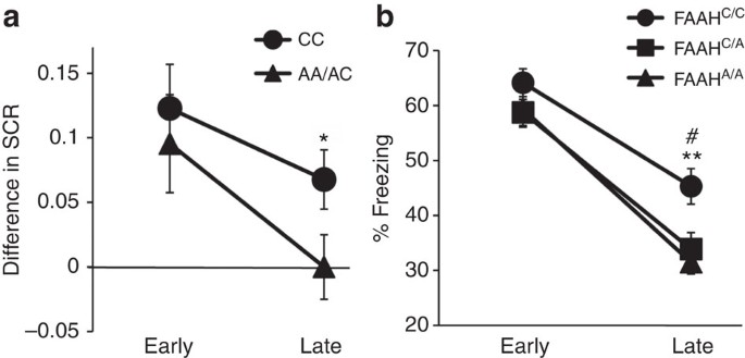
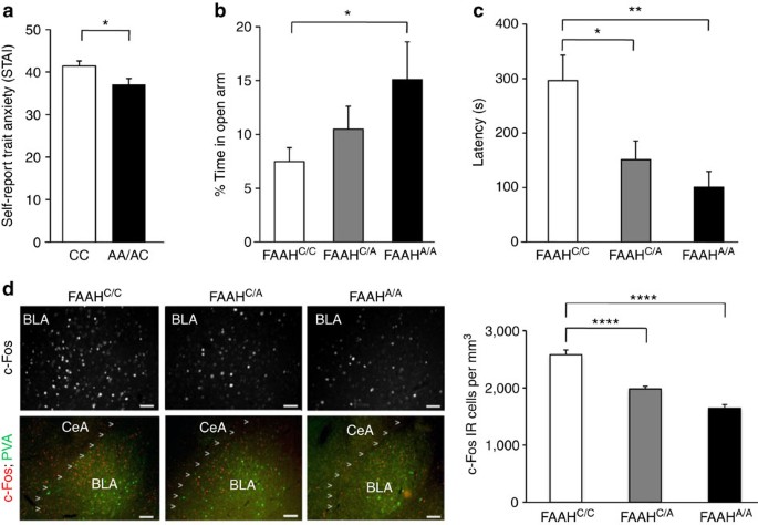
 

•  Vitamin D - I notice I can increase my serotonin to the point where I feel apathetic if I take 5000 I.U. a day. So taking normal doses purely for normal serotonin function.


•  Sulbutiamine - Is taken to increase B1 in the brain and body. I had a previous experience years ago which were akin amygdala tickling and being happy on life. Also I believe this will help my sleep apnea, and has helped most with my indigestion problems.


•  Lavender - To increase anandamide levels. Lavender is an FAAH & MAGL inhibitor up to 80%, thus increasing your natural endocannbinoids. Also a good hypnotic aid (as is 400mg theanine).


•  Cocoa Powder -  

1.  While there is very little anandamide in cacao, there are 2 important chemicals found in much higher concentrations in cacao that are more than likely the reason why we notice the mood elevating effects from cacao: N-oleolethanolamine (OEA), a known inhibitor of weight gain in mice, and N-linoleoylethanolamine (18:3 NAE), a known anti-inflammatory molecule — like anandamide — via the TRPV1 receptor). These two structural cousins of anandamide both inhibit the metabolic breakdown of anandamide and could potentially inhibit the breakdown of other cannabinoids including THC and CBD causing these compounds to stick around longer, providing even more benefits.

2.  One-week cocoa flavanol intake increases prefrontal cortex oxygenation at rest and during moderate-intensity exercise in normoxia and hypoxia. NEW & NOTEWORTHY For the first time, we showed that CF had beneficial effects on endothelial function at rest, as well as on prefrontal oxygenation at rest and during moderate-intensity exercise, both in normoxia and hypoxia. Moreover, we showed that CF intake inhibited oxidative stress during exhaustive exercise in hypoxia.

3.  Acute cocoa flavanols intake improves cerebral hemodynamics while maintaining brain activity and cognitive performance in moderate hypoxia. CF enhanced NVC in the right prefrontal cortex during several tasks (risk decision making, visual tracking, complex scanning, spatial orientation), while neuronal activity was not affected.

4.  In this study, the degree of cocoa alkalization caused a progressive, but not complete loss, of flavanol antioxidants, with about 40% retained in lightly dutched cocoas, 25% retained in medium dutched cocoas, and 10% retained in heavily dutched cocoas.

5.  Overall, when made with both water and milk the pure cocoa beverages (Green & Blacks and Cadbury Bournville) were shown to be richer in total polyphenols compared to lower purity cocoa powder varieties.


•  Anandamide research - 

1.  After it’s produced in the brain, anandamide is quickly broken down into other compounds by an enzyme called fatty acid amide hydrolase (FAAH). The slower FAAH works, the longer anandamide stays in the body.

2.  The research provides the first link between oxytocin  dubbed the "love hormone"  and anandamide, which has been called the "bliss molecule" for its role in activating cannabinoid receptors in brain cells to heighten motivation and happiness. A small number of neurons in the brain make oxytocin and use it as a neurotransmitter. When the scientists stimulated those neurons, they saw an increase in anandamide creation in the nucleus accumbens. More importantly, they found that blocking anandamide's effects also blocked the pro-social effects of oxytocin, which implies that oxytocin reinforces social ties by inducing anandamide formation. Our findings open the exciting possibility that drugs that block the degradation of anandamide, which are currently being tested for various anxiety disorders, could give a boost to the brain's own oxytocin and help people with autism socialize more," Piomelli said.

3.  Specifically, there is reduced FAAH expression associated with the variant allele that selectively enhances fronto-amygdala connectivity and fear extinction learning, and decreases anxiety-like behaviours.

4.  A Scottish woman with a rare genetic mutation in her FAAH gene with resultant elevated anandamide levels was reported to be immune to anxiety, unable to experience fear and insensitive to pain. The frequent burns and cuts she suffered due to her hypoalgesia healed quicker than average.

5.  Results showed that participants with PTSD, especially women, had more CB1 receptors in brain regions associated with fear and anxiety than volunteers without PTSD. The PTSD group also had lower levels of the neurotransmitter anandamide, an endocannabinoid that binds to CB1. If anandamide levels are too low, Dr. Neumeister explains, the brain compensates by increasing the number of CB1 receptors. "This helps the brain utilize the remaining endocannabinoids," he says.

6.  FAAH inhibition produced a 10-fold increase in baseline anandamide. This was associated with potentiated recall of fear extinction memory when tested 24 hours after extinction training. FAAH inhibition also attenuated autonomic stress reactivity, assessed via electrodermal activity, and protected against stress-induced negative affect, measured via facial electromyography. The beneficial effects of FAAH inhibition on fear extinction, as well as stress and affect-related behaviors, provide a strong rationale for developing this drug class as a treatment for posttraumatic stress disorder.

7.  The findings suggest that overactive corticotropin-releasing factor (CRF) signaling in this region produces a wide range of effects that override the stress-reducing capabilities of a major eCB called N-arachidonoylethanolamine (anandamide), turning chronic stress into unchecked, or pathological, anxiety. The researchers concluded that long-term dysregulation of CRF-FAAH mechanisms in the amygdala keeps anandamide from doing its job. Without anandamide to balance out the system, the brain is primed to react to stress.


•  ALCAR & Egg Yolks - Was taking both previously when I experienced my first frontal lobes pop, so I want to experiment with them again. ALCAR also previously caused me to have motivation in the gym. ALCARs side effects I remember were irritability and insomnia, I’m hoping to counter the irritability with DMAE. Some things I recently learnt about ALCAR which is why I might have experienced a mini pop only after a month of amygdala tickling using the feeling method:

1.  LAC can also increase the structural plasticity in the medial amygdala and reverse depressive-like behaviors in a chronic restraint-stress model.

2.  Oral LAC increased neuronal branching, where medial amygdala stellate neurons were more complex and longer.

3.  They conclude that the medial amygdala is identified in this study as a novel target of structural and functional remodeling by stressors and the new antidepressant candidate acetyl-L-carnitine. 

4.  Acetyl-L-carnitine levels were also lower among those patients reporting a childhood history of abuse, neglect, poverty or exposure to violence.

5.  To the extent that some vulnerability to depression is based on these early childhood experiences, LAC treatment could theoretically reverse that vulnerability in the long term in a fashion different from traditional ADs.

6.  Furthermore, CRS (chronic restraint stress) mice treated with LAC show resilience of the CRS-induced structural remodeling of medial amygdala (MeA) stellate neurons.

7.  Our findings identify MeA stellate neurons as an important component in the responses to stress and LAC action and show that LAC can promote structural plasticity of the MeA.


#179 Mr Serendipity

  • Topic Starter
  • Guest
  • 986 posts
  • 19
  • Location:UK
  • NO

Posted 07 March 2020 - 03:32 PM

OK a mini update:


1. Emotional flashbacks have been pretty non existent. Attribute this mainly to DMAE, and maybe egg yolks in the morning. Also I never posted this, but I did get hold of some 40mg of propranalol and would try to use it whenever I would get one, but most times I took it only after hours of remembering I had it. But I definitely think DMAE is the main thing at work, because the other day when I didn't take any of my supplements and clicked throughout the day, I was getting moody in the evening, this doesn't happen on DMAE, neither do emotional flashbacks.


2. I've split my DMAE to 1/4, so roughly 44mg (175mg pill / 4). At 1/4 dose I still experience emotional stability but with less of an emotional dully effect and less insomnia.


3. I can't take ALCAR, it completely screws me up, worst sleep ever, worst thinking energy ever. This was always a possibility for me ever since I became allergic/sensitive to coq10 and l-carnitine tartrate after 2015, when I was taking 600mg of coq10 and 3g of l-canitine tartrate a day and hitting the gym like a beast. ALCAR's definitely a supplement to look into for amygdala tickling and energy if you can take it.


4. I've increased my Vitamin D to 5000 I.U. from today. I remember experimenting this high before and noted it in this thread it increased my serotonin a lot making me apathetic. But now I'm finding I need something to combat the mental stimulation at the end of the day from everything else I've added since then, but also give me mental energy at the same time. It's hard to explain, but basically I'm hoping Vitamin D will contribute to motivational energy, but also calm me at the end of the day by increasing my serotonin and melatonin production.


5. I've drank cocoa for 26 days in a row now. My memory has improved significantly, I'm remembering memories from my life more and more each day. My memory was pretty bad before this, short term and long term, they'd be things my friends would say do you remember this or that event I wouldn't remember any of it. Anyway I mostly attribute my increase in memory to cocoa and amygdala tickling. I wrote a little bit more of this here: https://www.longecit...te/#entry886520


6. The other day I was very high on life and experiencing euphoria listening loudly and singing along to gospel music. 


7. I joined a new gym that just opened near me this week, it's a 24 hour one, which is going to help with my non normal sleeping routine. So I'm hoping to start getting 30 mins of cardio 4 times a week in again, lets increase those anadamide levels further and see if it can help me pop, and lose some weight (currently 109.2kg). 


8. It's been 19 days where I've stopped all visualization of amygdala tickling and only been using the feeling method. Pretty sure it works, but still require more time test out.


Summary:


Basically since the discovery of DMAE and it giving me emotional stability and preventing emotional flashbacks completely. Plus that daily cocoa drinking this past month. And the occasional euphoria. Well things are seriously looking up, not one of those times I thought the supplement was working and would then get another emotional flashback, so I'm optimistic, but need to give it a few more months. We can use this post as a marker, 7th of March, to see if I experience 1 emotional flashback from now on.


All Praise to God!


sponsored ad

  • Advert
Advertisements help to support the work of this non-profit organisation. To go ad-free join as a Member.

#180 Mr Serendipity

  • Topic Starter
  • Guest
  • 986 posts
  • 19
  • Location:UK
  • NO

Posted 12 March 2020 - 04:31 PM

Okay another update.


So I've been exercising regularly since I joined the new gym. Been 4 times in a week, 30 minutes on the cross trainer (elliptical), and getting around 90% of my maxiumum heart rate. Oh and I quit smoking.


But been feeling like shit. I mean terrible. Sleep has been majorly suffering, can't fall asleep, major sleep disturbance. 


This actually happened last time when I started exercising. It's like my body hates ketosis so much, doesn't matter if I restrict calories via diet, or burn them off, it gives me terrible sleep. This might be a connection also to my mitochondria, and how I can't take ALCAR or CoQ10 anymore.


Anyway back to yesterday. After a week of suffering from sleep and exercising. I decide to screw it and test a high dose of 5-htp, 200mg i think, but definitely 150mg.


Woke up today feeling normal, and not feeling overstimulated. I've been hesitant to take 5-htp regularly and long term (only using it at 50mg as an occasional sleep aid) because people have reported negative effects in the long term. However I don't know whether I have a choice, I think I have low serotonin.


I also read someone linked brain zaps with low serotonin, and was getting them last year at one point.


Basically serotonin had that dulling effect I'm sure everyone is aware of. But I think it's something I need to look into increasing, because I will so normal today, and this is without testing with DMAE.


Also I had a strange feeling while dreaming a couple of days ago when I took 5-htp at a lower dose, and I really wanted to write about it, felt like a feeling I hadn't felt in a long time, a different perspective and understanding. Then when I woke up, I lost it. But it was definitely related to amygdala tickling and the 5-htp I took.


Also I hypothesized serotonin could have been a factor in my case, because of the enhanced effects of amygdala tickling I got with weed, and how recreational drugs generally effect the serotonin system. 


Anyway it's very beneficial discovery to me, and I want to test more with high doses of 5-htp before bed. I just wake up so refreshed and normal. Things are getting to the end of my stack I can feel the gaps filling in. Just need to keep up with the cardio as well.


Edited by Jesus is King, 12 March 2020 - 04:32 PM.


Also tagged with one or more of these keywords: amygdala, visualization, frontal lobes, brain exercise

50 user(s) are reading this topic

0 members, 50 guests, 0 anonymous users